Author name: Vishi Sethi

Blog

Biofilm Formation and Gastrointestinal

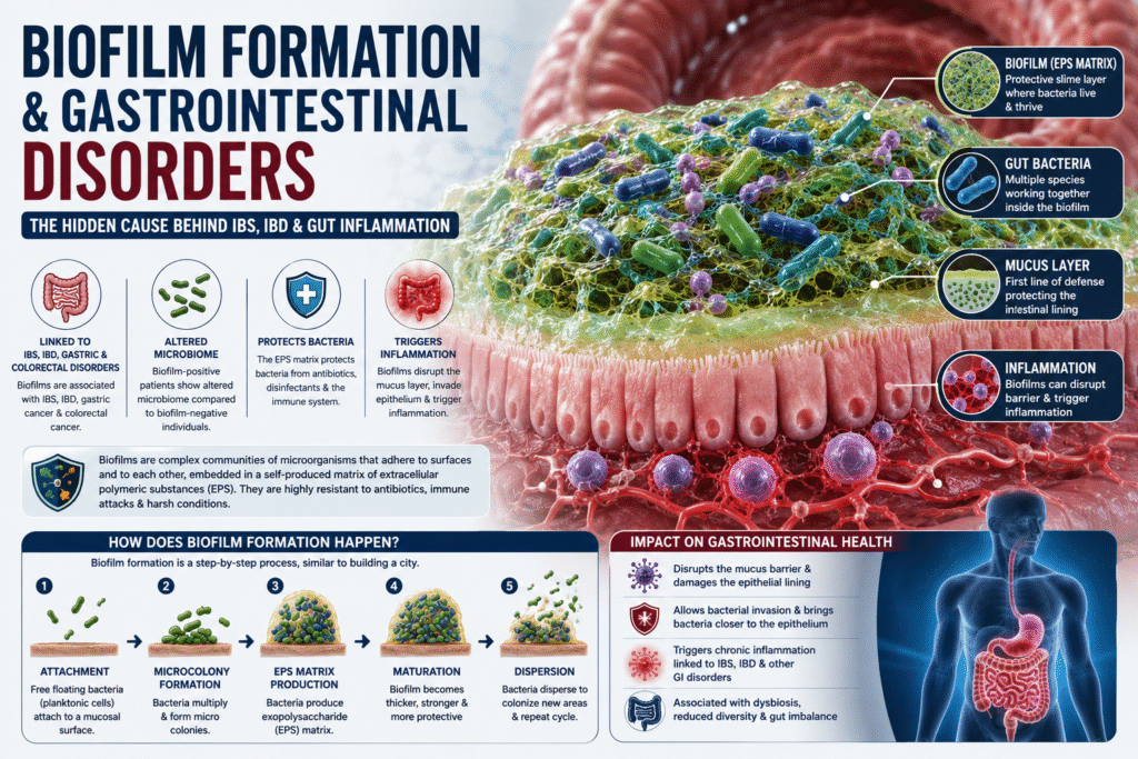
Biofilm Formation and Gastrointestinal Disorders As per the reports of the National Institute of Health (NIH), about 80% of human infections affecting the gastrointestinal, genitourinary (UTIs), respiratory systems, oral mucosa and teeth, eyes, middle ear and skin are caused due to BIOFILM FORMATION by biofilm-associated microorganisms. The ability to form biofilms is a universal attribute of bacteria, and biofilms play a role in several infections including – infection of indwelling medical devices, wound infections, bacterial carditis (heart-infection), otitis media (middle ear infection), dental carries, and lung infections of cystic fibrosis patients (13).  Gut microbiota dysbiosis, mucus disruption, and epithelial invasion are associated with pathogenic biofilms that have been linked to gastrointestinal disorders such as irritable bowel syndrome (IBS), Inflammatory Bowel Diseases (IBD), gastric cancer, and colorectal cancer (2). Intestinal biofilms are highly prevalent in ulcerative colitis and irritable bowel syndrome (IBS) patients, and most endoscopists have observed such biofilms during colonoscopy, in the gastrointestinal environment (GI tract) (2). IBS and IBD are the two most frequent GI disorders, together affecting >10% of the Western population. A recent clinical study revealed endoscopically visible mucosal biofilms in 57% of IBS, 34% of ulcerative colitis (UC), and 22% of Crohn’s disease (CD) patients (6% healthy; 976 patients in Austrian cohort and 450 in German cohort) (2,12). Biofilm-positive UC and IBS patients had an altered microbiome compared with biofilm-negative individuals, a finding independent of disease state (2.12).  Antibiotics and food additives might contribute to the reduced microbial diversity and biofilm formation, and it is plausible that food industrialization is linked to biofilm formation frequency, aligning with the higher prevalence of IBS and IBD observed in Western populations (2). What are Gastrointestinal Biofilms? Gastrointestinal biofilms are matrix-enclosed, highly heterogenic and spatially organized polymicrobial communities that can cover large areas in the gastrointestinal tract. The human gastrointestinal (GI) tract is the alimentary canal extending from the mouth to the anus and is the most densely inhabited environment of the human body. The GI tract harbors a profusion of microorganisms with different lifestyles called the gut microbiota, accounting for around 30% of the human microbiome. Along the GI tract, bacterial density increases, with the highest density in the colon (109–1011 bacteria/mL). The interplay of bacteria with the environment and the host affects the microbiota’s phenotypical occurrence and composition. Many gut microbes live as free-floating cells in the lumen, whereas others adapt higher-ordered structures termed biofilms (2,3,4,5) (as shown in the picture).   The mucus layer predominantly comprises dynamic mucin glycoprotein sheets coating the epithelial surface, forming the main barrier between the intestinal epithelium and luminal content (2,6). It effectively protects the host from digestive enzymes, acids, microbial by-products, food-associated toxins, pathogens, and microbial infiltration, preventing infection and inflammation (2,6,7). A compromised mucus layer or defects in mucus production can facilitate bacterial colonization and mucosal biofilm formation (2,6,8). Biofilm formation on the outer mucus layer can lead to mucosal invasion and bring bacteria close to the epithelium, an event that is disease-associated (9,10). Polymicrobial biofilms naturally grow throughout the gastrointestinal tract, both at the epithelial surface and in the lumen as mucin-attached and food particle-attached colonies.   In simple words – A biofilm is a complex multi-cultural community of microorganisms, such as bacteria, fungi, or algae, that stick to each other and often adhere to a surface within a slimy, self-produced matrix known as the extracellular polymeric substance (EPS) or “slime.” This matrix, made up of sugars, proteins, lipids, and DNA, protects the microbes and helps them survive in harsh conditions by providing a shield against antibiotics, disinfectants, and the host immune system.   How does Biofilm Formation Happen? Let’s liken the multispecies bacterial biofilm to a city – where bacteria settle selectively, limit settlements of new bacteria, store energy in exopolysaccharide, and transfer genetic material horizontally all for the good of the many (1). There are several steps that we must take to optimize our lives in a city. The first is to choose the city in which we will live, then we must select the neighborhood in the city that best suits our needs, and finally we must make our home amongst the homes of many others. Occasionally, when life in the city sours, we leave. The same steps occur in the formation of a bacterial biofilm as shown in the picture below (1). To give a larger picture – Free floating Bacteria (known as planktonic cell) attach to a mucosal surface, form microbial colonies, flagellin is reduced and create a hydro-gel like structure called exopolysaccharide or EPS matrix to protect themselves from host’s immune system and antibiotics. This EPS layer secretes extra polymeric cellular substances such as water, polysaccharides, lipids, proteins, and extracellular DNA resulting in a three-dimensional network, known as EPS matrix, that provides mechanical and chemical stability. The matrix protects against host defense mechanisms, mechanical forces of intestinal peristalsis, and antimicrobials through slow or incomplete drug penetration (1). The Life Cycle and Architecture of Gastrointestinal Biofilms include the following steps and mechanism:   Attachment – bacteria attach to the mucosal surface or aggregate (to each other or luminal content such as undigested food particles or host mucins) using surface-expressed adhesion proteins, flagella, and pili to form microcolonies (2). Development – The mucus-attached microcolonies start cell division and establish an EPS matrix known as biofilm matrix. The synthesis and secretion of EPS matrix, includes polysaccharides, proteins, lipids, and extracellular DNA (their functionality is described below). The EPS also comprise a significant amount of water that produces (together with the biopolymers) a hydrogel-like biofilm matrix. The EPS matrix fills the space between biofilm cells and holds cells together and anchors them to the mucosal surface, protecting the microbial community from environmental stresses, antibiotics, and immune responses (2). Maturation – Microbes proliferate, forming structured colonies with channels and pores that help transport nutrients and remove waste. This structure is sustained and protected by the EPS matrix, allowing the biofilm to persist in diverse environments. Persister cells form in nutrient-deficient areas, typically at the core of the mature
Blog

LEAKY GUT SYNDROME –

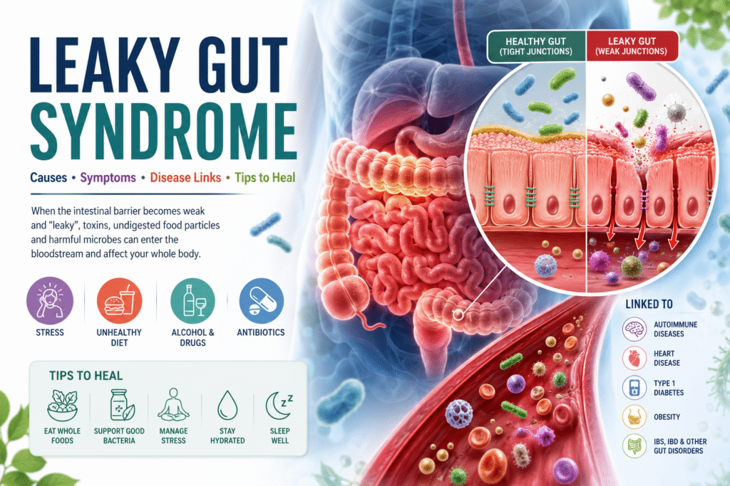
LEAKY GUT SYNDROME – Causes, Symptoms, Disease links and Tips to Heal The human body is in daily contact with potentially toxic and infectious substances in the gastrointestinal tract (GIT). The GIT protects the intestinal integrity by allowing the passage of beneficial agents and blocking the path of harmful substances.  Under normal conditions, a healthy intestinal barrier (gut lining) prevents toxic elements from entering the blood stream. However, factors such as stress, an unhealthy diet, excessive alcohol, antibiotics, and drug consumption can disturb the composition of the intestinal microbiota (gut flora) and homeostasis of the intestinal barrier of the intestine, leading to increased intestinal permeability.  The Intestinal Hy-permeability can allow the entry of harmful agents through the junctions of the intestinal epithelium, to leak into the blood stream and affect various organs and systems and is known as LEAKY GUT SYNDROME (LGS)! An increase in intestinal permeability is a sign of a disturbed intestinal barrier (2). According to the leaky gut syndrome (LGS) hypothesis, intestinal hyperpermeability may allow the entry of harmful microorganisms, toxins, or undigested food particles through the junctions of the intestinal epithelium, reaching the bloodstream and being able to affect the hormonal, immune, nervous, respiratory or reproductive systems (3). Thus, dysfunction of the intestinal epithelial barrier and increased permeability results in a “leaky gut” that is associated with intestinal disorders such as inflammatory bowel disease (IBD), irritable bowel syndrome (IBS), alcoholic liver disease (ALD), nonalcoholic fatty liver disease (NAFLD), steatohepatitis, liver cirrhosis, and collagen diseases (1). Leaky gut syndrome is also associated with extra-intestinal diseases (diseases that are not related to intestinal disorders) such as heart diseases, obesity, type 1 diabetes mellitus, and celiac disease (1).   Thus, the mucosal barrier is crucial to protect the body from exogenous harmful biological and chemical agents, such as microorganisms and environmental pollutants. The function of gastrointestinal epithelial barrier is to protect against the entry of foreign antigens and microorganisms, while allowing the absorption of essential nutrients, water and electrolytes (1,57).  The intestinal barrier consists of four components: microbial barrier, biochemical barrier, physical barrier and immune barrier (1,57). The microbial barrier is the intestinal microbiota, located in the lumen of an intestine. The microbiota produces many metabolically active compounds that show antimicrobial activity and affect the function of the entire intestinal barrier. Commensal bacteria digest certain food components and also compete with pathogens for nutrients (1,57). The biochemical barrier is mucus, which contains about 98% water (1) and, among others, mucins, glycoproteins, IgA antibodies, antimicrobial substances, produced by microorganisms—bacteria, viruses, fungi and intestinal cells. Mucus coats epithelial cells and protects them from the harmful effects of pathogenic microorganisms and toxic substances (57).  The physical (epithelial) barrier is an essential component of the entire intestinal barrier. It consists of a single layer of specialized cells: enterocytes, goblet cells (produce mucins), Paneth cells (produce antimicrobial peptides and proteins), enteroendocrine cells, M cells and intestinal stem cells. These cells undergo renewal every 3–5 days. Epithelial cells have a variety of functions and are closely interconnected (1,57).  The immune barrier is associated with the presence of lymphoid tissue in the intestines known as gut-associated lymphoid tissue (GALT). The GALT system is located in the mucosa and submucosa of the intestines, directly beneath the epithelial cells. This system consists of intraepithelial lymphocytes (IELs), Peyer’s patches, which are clustered lymphoid papules, and lymphocyte clusters. The GALT system has also been found to contain antigen-presenting cells (APCs), T lymphocytes, B lymphocytes, plasma cells, as well as macrophages, mast cells and dendritic cells (DC). The secretory IgA antibody (sIgA) is synthesized in the intestine in particular (1,57). This picture is taken from reference 1 This picture is taken from reference 57 The intestinal barrier is a selective barrier—its function is to allow the transport of digested food essential for the body’s function, but at the same time to keep harmful substances and microorganisms in the intestinal lumen, which requires strict regulation of the barrier’s permeability (1,57). This transport is regulated by tight junctions (TJ) (1,57). The TJ between enterocytes play a key role in providing an intestinal barrier. These junctions are composed of proteins, including occludin, claudin and junctional adhesion molecules (JAMs) and peripheral proteins known as zonula occludens (ZO-1, ZO-2, ZO-3), which bind to actin filaments (1). The primary role of zonulin is to dynamically open and close tight junctions between epithelial cells, thereby regulating paracellular permeability. When zonulin is released and its pathway is activated, it triggers intracellular signaling (including protein kinase C and cytoskeleton rearrangement) that leads to reversible disassembly of tight junction proteins such as ZO‑1 and occludin. Increased zonulin production has been observed under the presence of certain bacteria and food components, such as gliadin peptides found in gluten. Excessive release of zonulin results in weakening of TJ and consequent passage of antigens into the vicinity of immune cells and into the circulatory system. As a result, local inflammation develops and activated immune cells and cytokines can affect other organs or trigger immune-related diseases, e.g., autoimmunity (58). Elevated zonulin levels and increased permeability have been associated with several chronic inflammatory and autoimmune conditions, including celiac disease, type 1 diabetes, inflammatory bowel disease, and some neuroinflammatory and neurodegenerative disorders (59). Because of its strong association with barrier dysfunction, zonulin is being explored as a biomarker of impaired gut barrier function in various autoimmune and chronic inflammatory diseases. Pollution and climate change, chemical compounds commonly used in industry and households, ecosystem changes, unhealthy diet, and stimulants, mainly alcohol, tobacco and e-cigarettes, may disrupt the epithelial barriers of the skin and mucosal surfaces. Air, water and food pollution, microplastic particles, nanoparticles, household chemicals and tobacco smoke are the most common epithelial barrier disrupting factors (57). Therefore, pathogenesis of inflammatory bowel and leaky gut diseases is associated with multifactorial causes as discussed below.  Gut Microbiome and Leaky Gut The gut has more than 100 trillion bacteria (4), with an aggregate biomass of approximately 1.5 kg (5) composed of more than 200 microbial strains in an individual and

Blog

Exploring the Link Between Candida Overgrowth and Fatty Liver Disease

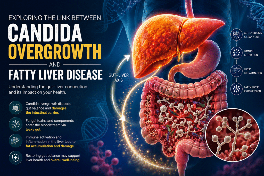
Exploring the Link Between Candida Overgrowth and Fatty Liver Disease What is a Fatty Liver Disease? Fatty liver disease, particularly Nonalcoholic Fatty Liver Disease (NAFLD), is a widespread metabolic condition impacting millions worldwide. Emerging research highlights not only the role of bacteria but also fungi, especially Candida species, in influencing liver health. This blog explores how Candida overgrowth may be linked to fatty liver and what it means for your health. Note – NAFLD is now called MASLD (Metabolic Dysfunction Associated Steatotic Liver Disease) as it is linked to metabolic issues like obesity, diabetes, and high cholesterol, characterized by fat buildup in the liver. What is Candida Overgrowth? Candida is a type of yeast that naturally lives in the gut, mouth, and other parts of the body. Under normal conditions, it coexists peacefully with the body’s microbial community. However, when Candida grows excessively—a condition known as Candida overgrowth—it can disrupt gut balance, potentially leading to digestive issues, immune dysfunction, and increased intestinal permeability (leaky gut). Interactions between intestinal fungal community (aka. mycobiome) and liver are anatomically and functionally bidirectional as explained below: Antigens derived from the gut commensal fungi can cross the gastrointestinal barrier and be transported via the portal vein to the liver, thus having an impact on its function. The immune cells in the liver can contribute to the host homeostasis through immune responses to the intestinal fungi (1,2). Thus, it has become apparent that gut mycobiome has a profound influence in modulating local as well as peripheral immune responses. However, like bacteria, fungi can be beneficial to host immunity. But they may also have deleterious effects (1,3) when the host immune system is compromised, or repeated rounds of antibiotics are used (4). This causes changes in the composition of the intestinal microbiota, promoting a series of liver diseases through the “entero-hepatic axis.”  Candida’s role in Fatty Liver Pathology Candida overgrowth is linked to fatty liver disease – alcoholic fatty liver disease -ALD (5) and non-alcoholic fatty liver disease – NAFLD (1) through mechanisms involving inflammation and immune response triggered by fungal components. A study (5) observed that alcohol-dependent patients displayed reduced intestinal fungal diversity and Candida overgrowth. Compared with healthy individuals and patients with non–alcohol-related cirrhosis, alcoholic cirrhosis patients had increased systemic exposure and immune response to mycobiota (1). Candida albicans and other fungi are found enriched in the gut microbiota of people with NAFLD, especially those with inflammation and advanced fibrosis (1). Inflammation is known to play a major role in the progression of NAFLD and NASH. Liver immune cells when exposed to fungal antigens and fungi derived metabolites elicit anti-inflammatory cytokines and chemokines, some of which can lead to liver damage (1,2). The fungal cell wall polysaccharide β-glucan can induce chronic liver inflammation by continuously activating the cellular inflammasome pathway leading to hepatocyte (liver cells) damage (1,5). Even though the association is evident, researchers caution that Candida may not directly cause fatty liver but that both conditions share overlapping mechanisms of gut barrier dysfunction and immune dysregulation as explained below: Gut Dysbiosis and Leaky Gut: Candida overgrowth alters the gut microbiome and damages the intestinal barrier. This allows fungal toxins, such as acetaldehyde and fungal cell wall components (like 1,3-β-glucan) to enter the bloodstream and reach the liver via the portal vein. Immune Activation and Liver Inflammation: The 1,3-β-glucan from fungal cell wall enters the liver via portal vein through the damaged intestinal mucosa and binds to CLEC7A on the surface of the liver Kupffer cell, then stimulates the Kupffer cell to secrete inflammatory cytokines like interleukin-1β (IL-1β). This immune activation causes liver inflammation, contributing to steatosis (fatty buildup) and liver cell injury (5,9). Clinically, many individuals with recurrent candida infections or overgrowth also have fatty liver issues, suggesting that candida-related gut dysbiosis and leaky gut can impair liver function and the immune system. Candida may also activate NF-kB-mediated inflammatory pathways affecting insulin resistance and lipid metabolism, which are key drivers in NAFLD development.  Research also shows increased systemic antibodies against Candida in patients with advanced fatty liver fibrosis. In this regard, Demir et al. examined the anti–C. albicans IgG antibodies in plasma samples of NAFLD and controls and found significantly higher IgG levels in NAFLD patients with advanced liver fibrosis. The authors assume that this probably indicates increased immune response to Candida albicans either due to the increased abundance of intestinal C. albicans or the relatively more frequent systemic exposure to C. Albicans (1,7). Toxin Production: Candida produces metabolites that may induce oxidative stress and exacerbate liver damage. It produces toxins such as acetaldehyde (9) that may cause systemic symptoms and candidalysin (6) that may promote alcohol associated liver disease. (see examples below) Metabolic Effects: Candida can worsen insulin resistance and lipid metabolism abnormalities, both of which are key drivers in the progression of fatty liver disease. Studies show that many people with Candida overgrowth also present with fatty liver or related symptoms like chronic fatigue, mental fogginess, and digestive disturbances. Chronic consumption of alcohol (ethanol) increases the fungal population and causes dysbiosis of the mycobiota in the intestine (right). The fungal dysbiosis results in higher amounts of β-glucan translocating across a damaged gut barrier to the liver (left). Increased β-glucan binds to CLEC7A on hepatic Kupffer cells and induces the expression and secretion of IL-1β. This cytokine contributes to ethanol-induced liver inflammation, hepatocyte injury, and steatosis. (Picture taken from reference 5) Association of Intestinal Fungi and Alcoholic liver disease (ALD) Chronic alcohol consumption is a well-known factor of increased intestinal permeability, and changes in the intestinal microbiota composition which may contribute to the development of alcohol related liver disease. Liver acts as a metabolic site for alcohol, when the body excessively consumes alcohol for a long time and exceeds the metabolic load of the liver, it can cause liver damage through multiple routes, and constantly develop into alcoholic fatty liver, alcoholic hepatitis, alcoholic cirrhosis and even liver cancer (9,10,11).   In ALD, increased ethanol and its metabolite acetaldehyde in the intestinal lumen cause weakening of intestinal tight junctions. Consequently, increased translocation of microbial-associated

Blog

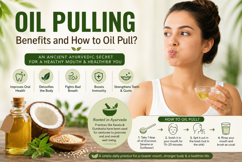
OIL PULLING – Benefits and How to Oil Pull?

OIL PULLING – Benefits and How to Oil Pull? It is a common belief that oral health serves as a gateway to general health. This implies that oral health significantly impacts the general health and wellbeing of an individual.  Dental diseases have detrimental effects on the functionality and quality of life of individuals. In addition, a strong relationship has been established between various oral and systemic diseases. In fact, the prevention and treatment of dental caries and periodontal disease have been shown to reduce the risk of diabetes and heart disease significantly. The use of oil pulling can be frequently found in ancient medical text and is supported by recent studies for its efficacy and long-term use for maintaining and improving oral health.  The oral cavity serves as a focal point of entry for pathogens into the systemic circulation. While the host immune system of a healthy individual prevents the body from virulent microorganisms, a breach in the physical barriers in the oral cavity may provide access to into the systemic circulation. Similarly, a lack of oral hygiene allows an increase in virulent microbial colonization of the oral biofilm. Therefore, mechanical and chemical means of controlling the quantity and quality (virulence) of the oral biofilm is important in preventing systemic diseases and particularly periodontal diseases such as gingivitis and periodontitis (1). Oil is pulling or oil swishing is the ayurvedic way of maintaining oral health and improving overall immune system. Oil pulling also acts as an excellent detoxifying agent in healing the body inside. It is a widely accepted fact that most of the diseases start because of the unhealthy mouth. Most of the chronic illness are directly related to an unhealthy mouth like gum disease or tooth decay. As per Ayurveda – Oil pulling is incredibly effective in brightening teeth, healing gums, preventing bad breath, quenching inflammation, and healing oral infections. For example – Brushing is contra indicated in the cases of mouth ulcer, fever, indigestion, those who have tendency to vomit, asthma, cough, thirst. Oil pulling can be used to clean the oral cavity in all these cases (2). Gundusha and Kavala Graha are two primary oral cleansing techniques used in Ayurveda; as a specialized therapy to treat as well as to prevent oral diseases. Gundusha involves filling the mouth completely with fluid so that gargling is impossible. In Gundush, the oral cavity is filled completely with liquid medicine, held for about 3-5 minutes, and then released. In Kavala Graha, a comfortable amount of fluid is retained with the mouth closed for about 3 minutes and then gargled. It is a simple rejuvenating treatment, which, when done routinely, enhances the senses, maintains clarity, brings about a feeling of freshness, and invigorates the mind. These oral cleansing techniques can also benefit bad breath, dry face, dull senses, exhaustion, anorexia, loss of taste, impaired vision, sore throat, and all kapha related imbalances (9). This article – highlights the importance of incorporating oil pulling as a component of daily oral hygiene which can significantly improve oral and general health. What is Oil Pulling? Oil pulling, in CAM (Complementary and Alternative Medicine), is a technique that involves vigorous swishing of oil in the mouth, to achieve oral and systemic health benefits, like the modern-day use of mouthwashes and oral rinses. It is a powerful detoxifying Ayurvedic technique that has recently become very popular as a CAM remedy for many different health ailments. In the Ayurvedic text “Charaka Samhita,” it is mentioned as Kavala or Gundusha and is claimed to cure about 30 systemic diseases ranging from headache, migraine to diabetes and asthma (1). It has been used for centuries for the treatment and prevention of various oral and systemic diseases, using edible oils derived from either sunflower, sesame, or coconut. Oil pulling has been used extensively as a traditional Indian folk remedy for many years to prevent tooth decay, oral malodor (bad breath), bleeding gums, dryness of throat, cracked lips and for strengthening teeth, gums and the jaw (1).  In addition to brushing your teeth, flossing, and scraping your tongue, oil pulling with sesame or coconut oil is a safe and effective bonus to a healthy oral hygiene routine. It helps in rebalancing oral microbiome and improving oral and dental health. Ayurveda advises oil gargling to purify the entire system; as it holds that each section of the tongue is connected to different organ such as to the kidneys, lungs, liver, heart, small intestines, stomach, colon, and spine, similarly to reflexology and TCM (9).  Scientific evidence suggests that oil pulling therapy may reduce the total oral bacterial count and reduce plaque and gingival scores. Furthermore, it has also shown to diminish the susceptibility to dental caries from marked to slight or moderate level (1).  How does Oil Pulling work? (Mechanism of action) Both Western medicine and Ayurveda use the tongue as an important diagnostic tool, indicating that a healthy mouth and a healthy tongue are interrelated with the health of the entire body. Thus, supporting our oral hygiene and is a benefit for both our dental and general health (3). In fact, our mouths host over 600 different species of bacteria that populate the teeth, tongue, soft tissues of the cheeks and palates, and our tonsils. The oral cavity further adjoins the esophagus, nasal passages, sinuses, and the intricate ear cavities. You can see why bacteria in the mouth is a big deal! (3) Many of these bacteria are necessary for a healthy oral microbiome, but some, such as Streptococcus mutans, can cause problems if left unchecked—tooth decay, bad breath, gingivitis, and strep throat, to name a few. Poor oral hygiene can allow harmful bacteria to flourish, leading to various oral health issues (1, 3). Bacteria are single-celled organisms, enclosed by a lipid membrane. These bacteria in the mouth are attracted to the lipid structure of the oil, pulled from the oral tissue by adhering to the fat molecules of the oil, then flushed away through the act of swishing oil and spitting it out. This process helps in

Blog

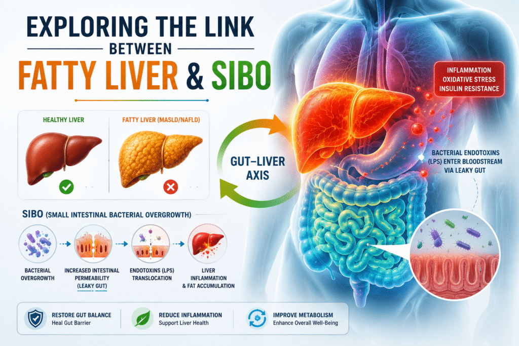
EXPLORING THE LINK BETWEEN FATTY LIVER AND SIBO

EXPLORING THE LINK BETWEEN FATTY LIVER AND SIBO Fatty liver disease, particularly Nonalcoholic Fatty Liver Disease (NAFLD), now known as Metabolic Dysfunction-Associated Steatotic Liver Disease (MASLD), has become a major global health concern. In recent years, MASLD has been the second leading cause of end-stage liver disease worldwide (28).  Increasing evidence reveals a fascinating connection between NAFLD and gut health, specifically a condition known as Small Intestinal Bacterial Overgrowth (SIBO). The correlation between small intestinal bacterial overgrowth (SIBO) and nonalcoholic fatty liver disease (NAFLD) has gained heightened acknowledgment, especially in the late phases of liver disease. Today, we explore the scientific links between these two conditions and why understanding this relationship matters. Understanding The Gut-Liver Axis The gut epithelium is a natural barrier that selects entry of useful substances present in the lumen, as nutrients, and keeps at bay bacteria, their bio-products and other potentially harmful elements. Tight junctions, specialized intercellular structures, assist this control. Derangement of the homeostasis between bacteria and the host, as occurs in SIBO (enhanced amount and/or changes in the type of bacteria in the gastrointestinal tract), may cause disruption of the intercellular tight junctions and subsequent increase in intestinal permeability leading to bacterial translocation (BT), i.e., transportation of bacteria and bacterial products from the intestinal lumen into the blood (4). The portal vein and the hepatic artery supply blood to the liver. The portal blood contains products of digestion and microbial products derived from the gut microbiota. This blood is carried to the liver. Therefore, Liver is the first site of exposure and filtration that consists of microbial products from the gut, such as LPS, lipopeptides, unmethylated DNA, and double-stranded RNA, which may evoke inflammatory reaction contributing to the progression of the liver disorder (4). This bidirectional relationship of the gut ecosystem and liver is imperative both physiologically and pathologically. Generally, the liver receives rich nutrients, microbial metabolites, and subproducts from the intestine and secretes bile into the small intestine (5). An integrated gut barrier also protects against toxins to maintain internal homeostasis.  This gut-liver axis is regulated and stabilized by a complex network of metabolic, immune, and neurosecretory interactions between the gut, microbiota, and liver. Disruption of this equilibrium may lead to gut dysbiosis and liver injury (5). SIBO as we know is the clinical manifestation of gut microbial dysbiosis. Therefore, the bidirectional relationship between Small Intestinal bacterial overgrowth (SIBO) and fatty liver disease, particularly non-alcoholic fatty liver disease (NAFLD), is characterized by mutual influences through gut-liver axis dysfunction, inflammation, and metabolic disturbances.  Association between SIBO and Fatty Liver Small intestinal bacterial overgrowth (SIBO) is a condition marked by excessive growth of microbes in the small intestine, resulting in various digestive issues including bloating, satiety, and malabsorption. In healthy people, the small bowel has a relatively low bacterial concentration, around 103–104 colony-forming units per milliliter (CFU/mL) (1,6). However, when this balance is disrupted, bacteria from the colon or oral cavity can colonize the small intestine, resulting in SIBO. Factors contributing to this condition include reduced gastric acid production, impaired intestinal motility, insufficient production of bile and dysfunction of the ileocecal valve (1,7). It may present in a range of symptoms, from moderate pain to severe nutritional deficiencies, weight loss, and shortages in crucial minerals and vitamins, including vit B12, A, D, E, iron, choline, calcium, fats, carbohydrates, protein and bile salt deconjugation (1,8). However, it has been shown that intestinal dysbiosis, endotoxemia (bacterial toxins in blood) and bacterial translocation may contribute to inflammation and Insulin Resistance (3,9,10,11,12). This directly seems to disrupt the functioning of the gut–liver axis, which may influence the incidence and progression of NAFLD (3,13). Non-alcoholic fatty liver Disease is the most frequent cause of chronic liver sickness globally, with a spectrum spanning from simple steatosis to inflammation of the hepatocytes, fibrosis, cirrhosis, and even hepatocellular carcinoma (1,14). The link between SIBO and nonalcoholic fatty liver disease (NAFLD) has attracted increased attention since studies show that the gut-liver axis plays a significant role in the pathophysiology of steatosis liver disease (1,15). The transfer of bacterial metabolites from the stomach to the liver may promote scarring and inflammation, thereby aggravating liver damage (1,16).  How SIBO affects Fatty Liver? 1.Inflammation – SIBO leads to an overgrowth of bacteria in the small intestine, causing increased intestinal permeability “known as leaky  gut or gut barrier dysfunction.” This allows bacterial endotoxins, especially lipopolysaccharides (LPS), to enter the bloodstream and reach  the liver via the portal vein, triggering chronic liver inflammation. These endotoxins and bacterial products activate inflammatory pathways (immune responses) in the liver, releasing proinflammatory cytokines (e.g. TNF-α, IL-6) which promote fat accumulation, insulin resistance, liver inflammation, fibrosis, and disease progression in NAFLD. Example – It has been demonstrated in animal models that a four-week HFD (high fat diet) increases LPS contained in the gut microbiota and plasma LPS concentrations two to three times, which is considered metabolic endotoxemia. The induction of metabolic endotoxemia in mice, by continuous subcutaneous infusion of LPS for four weeks, was followed by a rise in the following parameters: fasting glycemia, insulinemia, markers of inflammation, liver triglyceride content, liver insulin resistance, and whole body, liver and adipose tissue weight gain in a similar amount as occurred in HFD fed mice (4,17).  Large amount of fructose consumption is also related to increase in endotoxin serum levels, proinflammatory response and steatosis. It was demonstrated in an elegant study conducted by Bergheim et al. (4,18) that mice fed with fructose showed increased endotoxin levels in the portal blood, and higher intrahepatic lipid accumulation, lipid peroxidation and TNF-α expression.   2.Metabolic disruption – SIBO also disrupts bile acid metabolism, nutrient absorption, and adipose tissue function, contributing further to liver fat deposition and dysfunction.  Bile acids are synthesized in the liver from cholesterol, conjugated (joined) with glycine or taurine, and secreted into the small intestine, where they help digest fats and maintain gut microbial balance through their antimicrobial properties (3). In a healthy gut, most bile acids are absorbed in the ileum, while the rest reach the colon, where

Blog

THE KAPHA BODY TYPE

THE KAPHA BODY TYPE According to Ayurveda, each one of us are born with a certain body type or constitution known as Prakruti. When we deviate from our natural constitution (due to stress, unhealthy diet, poor lifestyle, medicines etc.), we attract diseases. The diseased state or an imbalance in our energies is known as Dosha or Vikruti.   In Ayurveda there are 3 types of Doshas or body types – Vata, Pitta and Kapha. In this article we are going to discuss the Kapha Dosha! Our body is made up of 5 elements air, water, earth, fire and ether.  The Kapha dosha – is principally a combination of Earth and Water elements. It is the energy that forms the body’s structure and provides the “glue” or cohesion that holds the cells together, lubricates joints, moisturizes the skin and maintains immunity. It governs the growth of the dhatus (bodily tissues) – the blood, fat, muscles, bones, marrow and other fluids in the body. It is also responsible for healing. The qualities of Kapha dosha are – moist, cold, heavy, slow/dull, soft, sticky or cloudy, oily, liquid, slimy/smooth, dense and static. In Ayurveda, there is a law which states that “like increases like and opposites decrease.” When similar qualities come together, their quantitative expression increases. For example, Winter season has attributes of liquid, heavy, cold, sticky and cloudy. Therefore, in the winter, when these characteristics predominate in the external environment, internal kapha tends to be increased.  Physiologically, kapha moistens food, gives bulk to our tissues, lubricates joints, stores energy, and relates to cool bodily fluids such as water, mucous, and lymph. Psychologically, when in balance – Kapha is expressed as love, calmness, and forgiveness. When out of balance – it leads to attachment, greed, possessiveness, and congestion disorders. The main seat of kapha in the body is stomach and chest. It is also located in throat, lungs, head, lymph, fatty tissue, connective tissue, ligaments, and tendons. Why the “Seat” Matters? Understanding the seat of a dosha helps in its treatment. The Chest connection: Since Kapha sits in the chest, breathing exercises (Pranayama) and heart-opening yoga poses are the fastest ways to “stir” and clear excess Kapha. The Digestive connection: Even though it sits in the chest, Kapha accumulates in the stomach first. This is why a “sluggish stomach” is often the first sign of a Kapha imbalance before it moves up into the lungs as congestion. Common Kapha Disorders of the Body Obesity, Diabetes, Colds and Flu, Yeast conditions, Sinus congestion, Anorexia and bulimia, lymphatic system disorders, excessive sleeping, water retention/bloating, allergies, excess phlegm and mucous conditions, intolerance of cold and damp, asthma, low thyroid function, heart disease. Characteristics of the Kapha Individual The phrase Kapha comes from two Sanskrit roots, ‘ka’ meaning water and ‘pha’ meaning to flourish – that which is flourished by water. Kapha’s nature is also Earth, so Earth and Water give kapha individual its definitive qualities. Qualities received by kapha from earth include heaviness and stability, and from water include oiliness and smoothness. Therefore, Kapha molecules tend to stick together to form dense masses (earth element) and give the body a chubby shape.  Physiological Characteristics of Kapha person (The Kapha Body) Because Kapha is naturally heavy, slow, and cool, its physiological traits reflect a “solid” and “enduring” nature. Physical Build & Structure Frame: Generally broad-shouldered with a sturdy, “large-boned” skeletal structure. Kapha types are naturally strong and have the highest physical endurance of all three doshas. Weight: A natural tendency to gain weight easily and a significant challenge in losing it. This is due to a slower basal metabolic rate. Joints: Well-lubricated, large, and hidden (not prominent or “knobby” like Vata). They rarely suffer from “popping” joints but may experience fluid retention or stiffness. Skin, Hair, and Nails Skin: Typically thick, pale, and cool to the touch. It is naturally oily and well-hydrated, which often leads to fewer wrinkles and a more youthful appearance as they age. Hair: Thick, voluminous, and often wavy or curly. It tends to be lustrous and strong, though it can become oily if Kapha is imbalanced. Eyes: Large, attractive, and “liquid” or soft in appearance. They usually have thick, long eyelashes and clear, white sclera (the whites of the eyes). Digestion and Metabolism Appetite: Regular but slow. Kapha types can often skip a meal without feeling irritable (unlike Pittas), as their bodies are excellent at storing energy. Metabolism (Agni): Known as Mandagni (slow/dull fire). Digestion takes a long time, and they may feel heavy or sleepy immediately after eating. Elimination: Bowel movements are usually regular, soft, and oily. Sleep and Energy Levels Sleep: Deep, heavy, and long. Kaphas are the “heavy sleepers” of the world and often find it very difficult to wake up in the morning. Stamina: While they are slow to start (inertia), they have incredible long-term stamina. They can work or exercise for long periods once they get moving. Pulse: The Kapha pulse is described as “Swan-like” (Gaja Gamini). it is slow, steady, and regular. Feature Characteristic Complexion Pale, fair, uniform Body Temp Cool and slightly damp (clammy hands) Voice Deep, pleasant, and resonant Immunity Naturally strong; best resistance to disease Tendencies Excess mucus, congestion, and water retention Psychological Characteristics of Kapha person (The Kapha Mind) Qualities of Balanced Kapha Individuals Loving, Compassionate, Patient, Sweet, Forgiving, Gentle, Emotionally Stable, Inherent Desire to Help Others, Calm, Loyal, Nurturing, Accepting of Others, Strong Stamina and Endurance, Romantic, Sensual, endowed with excellent long-term memory, long term thinking and planning, good listeners, deeply satisfied with life and good natured Qualities of Imbalanced Kapha Individuals Greedy, unchanging – rigid, Hoard material things, Unable to say No, Easily attached to people and things, Easily taken advantage of, Possessive, Take on other people’s problems, fearful of letting go, overly passive, lethargic, Unable to express thoughts and emotion, envious, give up easily, slow to understand/grasp things, depressed for long periods of time, introverted, complacent, living life on the surface Kapha dreams – One of the important functions

Blog

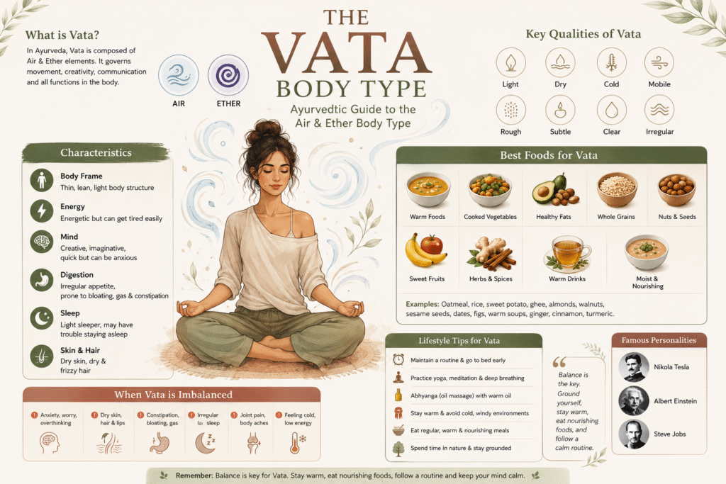
THE VATA BODY TYPE

THE VATA BODY TYPE According to Ayurveda, each one of us are born with a certain body constitution known as Prakruti. When we deviate from our natural constitution (due to stress, unhealthy diet, poor lifestyle, medicines etc.), we attract diseases. The diseased state or an imbalance in our energies is known as Dosha or Vikruti.  In Ayurveda there are 3 types of Doshas or Body types – Vata, Pitta and Kapha. In this article we are going to discuss the Vata Dosha! Our body is made up of 5 elements air, water, earth, fire and ether.  Vata dosha – is principally a combination of space and air elements.  The Sanskrit term Vata is related to the verb “vah,” meaning vehicle, to carry or to move which translates as “wind’ or “that which moves things.” So, Vata is the energy of the movement and the force governing all biological activity. It is the principle of mobility that regulates all activity in the body, from how many thoughts one might have during a given period to how efficiently food moves through the intestines. The Vata quality is responsible for joy, happiness, creativity, speech, sneezing, and elimination, to name just a few functions. Vata is often called the “King of the Doshas” since it governs the body’s greater life force – “Prana” and “gives motion to Pitta and Kapha.” Thus, when Vata (Prana) leaves the body, life ceases. The qualities of Vata dosha are – dry, rough, light, mobile, clear, subtle, and cold. A Vata individual will display physical and mental characteristics that reflect these qualities in both balanced and imbalanced state. Physiologically, Vata governs anything related to movement, such as breathing, talking, nerve impulses, movements in the muscles and tissues, circulation, assimilation of food, elimination, urination, and menstruation.  Psychologically, Vata governs communication, creativity, flexibility, and quickness of thought. The main seat of Vata in the body is colon. It is also located in thighs, bones, joints, ears, skin, brain, and nerve tissues.  Why Knowing the “Seat” Matters? Understanding the seat of a dosha helps in its treatment. In Ayurvedic medicine, if you want to calm a restless mind or soothe “windy” joints, you don’t just treat the head or the knees—you treat the colon. This is why: Digestion First: Keeping the colon hydrated and moving (avoiding constipation) is the #1 way to keep Vata balanced. Warmth & Oil: Since Vata’s seats are prone to cold and dryness, applying warm oils (Abhyanga) to the skin and hips is highly effective. Grounding: Focusing on the lower half of the body through grounding exercises helps “descend” the flighty energy of Vata back to its seat. Characteristics of the Vata Individual In Ayurveda, a Vata individual is defined by the elements of Air and Space. Their physiology is characterized by lightness, dryness, coldness, and irregularity. Because Vata governs all movement in the body, a Vata person’s system is often active but easily exhausted. The Vata Paradox: While they are the most energetic and creative of the types, their physiology is the most fragile. They are like a high-performance sports car with a very small fuel tank. Physiological Characteristics of Vata person (The Vata Body) General Build and Frame Structure: Vata types are usually at the extremes—either very tall or very short. They have a naturally thin, slender, or “willowy” frame. Weight: They generally have a low body weight and find it very difficult to gain weight and even harder to keep it on. Their metabolism is fast but erratic. Bone Structure: Because they lack subcutaneous fat, their joints, veins, and tendons are often very prominent or “knobby.” Bones and joints are often prominent or “bony” due to a lack of padding. It is common for their joints to make cracking or popping sounds. Skin, Hair, and Nails Skin: Naturally dry, thin, and cool to the touch. They are the most prone to chapping, cracking, and premature wrinkles. Veins are often visible beneath the surface. Hair: Hair is often thin, frizzy, or curly and prone to being brittle. It tends to be dry and can be prone to split ends.  Nails: Often brittle, thin, and prone to breaking or peeling. Face and Senses Face: Face is often thin and long with small, deep-set, or active eyes. Eyes: Usually small, deep-set, and active (sometimes described as “shifty” or “darting”). They may lack the luster or moisture seen in other types. Teeth: Can be irregular, protruding, or large. Digestion and Metabolism (The “Variable” Nature) Appetite: Highly erratic. They may be ravenous one day and forget to eat the next. Digestion: Prone to “windy” digestive issues such as gas, bloating, and constipation. Their “Agni” (digestive fire) is like a candle in the wind—unstable and easily blown out. Excretion: Bowel movements are often dry, hard, and irregular. Energy and Movement Activity Level: They move quickly and talk fast. However, they have low endurance. They are “sprinters, not marathoners.” They tend to use up their energy in quick bursts and often experience a “crash” after periods of high activity. Sleep: Light and easily disturbed. Vata types are the most likely to suffer from insomnia or wake up feeling unrefreshed between 2:00 AM and 6:00 AM (the Vata time of night). Elimination: The “Seat of Vata” (the colon) tends toward dryness, making constipation a common physiological trait. Feature Characteristic Body Weight Low; difficult to gain weight Skin Texture Dry, rough, cool Joints Prominent; prone to cracking Hands/Feet Usually cold (poor circulation) Speech Fast, talkative, sometimes scattered Weather Preference Loves the sun; hates cold and wind Psychological Characteristics of Vata person (The Vata Mind) In balance – Vata individuals are dynamic, just like the wind at their foundation. These individuals experience life “below the surface” and have the natural ability to inspire others.  While Vata types may be inclined to switch careers throughout their lifetimes, they naturally make good teachers, artists, musicians, consultants, counselors, healing art practitioners, philosophers, foreign ministers, religious ministers, and are also suited to numerous other professions requiring creativity and

Blog

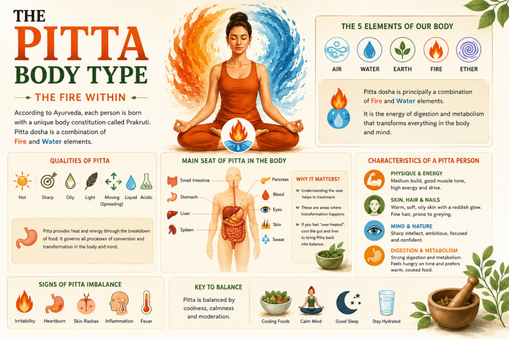
THE PITTA BODY TYPE

THE PITTA BODY TYPE According to Ayurveda, each one of us are born with a certain body constitution known as Prakruti. When we deviate from our natural constitution (due to stress, unhealthy diet, poor lifestyle, medicines etc.), we attract diseases. The diseased state or an imbalance in our energies is known as Dosha or Vikruti.  In Ayurveda there are 3 types of Doshas or body types – Vata, Pitta and Kapha. In this article we are going to discuss the Pitta Dosha!    Our body is made up of 5 elements air, water, earth, fire and ether.  Pitta dosha – is principally a combination of Fire and Water elements. The word Pitta is derived from the Sanskrit word “tap” meaning to heat and to be austere; and “translates as that which cooks.” It is the energy of digestion and metabolism in the body that functions through carrier substances such as organic acids, hormones, enzymes, bile and even the neurotransmitters and neuropeptides involved in thinking. Literally everything that enters the body needs to be “digested or cooked” from the sight of a full moon to a strawberry popped into the mouth. Some of pitta’s responsibilities are regulating the body heat through chemical transformation of food and giving a person appetite, vitality, learning and understanding.  The qualities of Pitta are – Hot, sharp, oily, light, moving or spreading, liquid and acidic. A pitta individual will display physical and mental characteristics that reflect these qualities in both balanced and imbalanced state.  Physiologically, Pitta provides the body with heat and energy through the breakdown of complex food molecules. It governs all processes related to conversion and transformation throughout the mind and body.  Psychologically, Pitta governs joy, courage, willpower, anger, jealousy, and mental perception. It also provides the radiant light of the intellect.  The main seat of Pitta in the body is the small intestine. It’s also located in stomach, liver, spleen, pancreas, blood, eyes, skin and sweat.  Why Knowing the “Seat” Matters?    Understanding the seat of a dosha helps in its treatment. Pitta is composed of Fire and Water elements. You’ll notice all its “seats  ” are areas where transformation happens: In the gut, food becomes energy. In the liver, toxins are processed. In the eyes, light becomes sight. In the skin, sunlight is synthesized and heat is released. Pro-Tip: If you feel “over-heated” (think heartburn, skin rashes, or irritability), Ayurveda suggests cooling these specific areas—especially the gut and the liver—to bring Pitta back into balance. The “Basement” Analogy: Think of the seats of the doshas like the utility rooms of a house. Vata is in the basement (colon), Pitta is on the ground floor (small intestine), and Kapha is in the attic (chest/head). If there’s a fire on the ground floor, you don’t run to the attic with a fire extinguisher—you go straight to the kitchen. Characteristics of a Pitta Individual    A Pitta individual is defined by the Fire and Water elements. Just like a steady flame, they are intense, transformative, and sharp. When balanced, they are brilliant leaders and “doers”; when out of balance, they can be a bit… combustible.  The balanced Pitta individual is blessed with a joyful disposition, a sharp intellect, and tremendous courage and drive. As the fire of the mind and body becomes unruly, however, the laughing Pitta quickly becomes the yelling – Pitta. Anger, rage, and ego replace Pitta’s positive attributes, leaving an individual who is bitter with life, and overbearing towards others. There is a saying that imbalanced Pitta individuals don’t go to hell; they simply create it wherever they go!  Pitta imbalances commonly manifest in the body as infection, inflammation, rashes, ulcers, heartburn and fever.  Physiological Characteristics of Pitta Person (The Pitta Body)    Physical Build and Energy Frame: Medium build with good muscle tone. They gain muscle more easily than Vata types but aren’t as naturally heavy-set as Kapha types. Physical Strength: They have moderate physical strength but high intensity. They often push themselves to the point of overheating during exercise. Sleep: They generally sleep soundly for moderate durations (6–7 hours). However, if their mind is overactive with a project, they may struggle to “turn off” the internal light. Skin, Hair and Nails Skin: The skin is typically warm, soft, and oily. It often has a reddish or pinkish undertone. They are prone to freckles, moles, and “hot” skin conditions like rashes, acne, or hives. Hair: Fine and silky; tends toward premature greying or thinning. Nails: Pink, soft, and flexible; typically, well-shaped and lustrous. Face and Senses Eyes: Pitta eyes are often very bright and sensitive to light. They frequently have a “sharp” or penetrating gaze. Many Pitta individuals end up needing glasses for nearsightedness due to the “heat” taxing the visual organ. Face: Heart-shaped or angular with sharp features (pointed nose/chin) and a glowing, reddish complexion. Senses: Sharp and acute; they have a strong sense of taste and a highly discerning “critical” eye. Digestion and Metabolism (The Powerhouse) The most defining physiological trait of a Pitta person is a strong, sharp appetite. Strong Digestion: They can typically digest almost anything, but they require regular meals. Their blood sugar tends to drop quickly if they don’t eat. Elimination: They usually have frequent, soft, or loose bowel movements. Because of their internal heat, they rarely struggle with constipation but may deal with “burning” during elimination if they eat spicy food. Cravings: They naturally gravitate toward cold drinks and sweet, cooling foods to balance their internal furnace. Thermoregulation and Energy Pitta types are “walking heaters.” Their bodies are constantly working to dissipate metabolic warmth. Body Temperature: Their hands and feet are usually warm to the touch, even in winter. Perspiration: They sweat easily and profusely, often with a strong or “sour” odor/fleshy smell. Energy level – Drive: High, focused, and steady energy. They are “doers” who push themselves until a task is finished. Endurance: Moderate to strong, but they tend to overheat. They are most energetic in cooler weather or during the early

Blog

Small Intestinal Bacterial Overgrowth (SIBO) –Symptoms, Subtypes, and Treatment

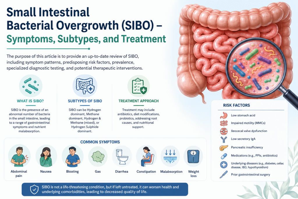
Small Intestinal Bacterial Overgrowth (SIBO) – Symptoms, Subtypes, and Treatment The purpose of this article is to provide an up-to-date review of SIBO, including symptom patterns, predisposing risk factors, prevalence, specialized diagnostic testing, and potential therapeutic interventions. What is SIBO? Small intestinal bacterial overgrowth (SIBO) is characterized by the presence of an abnormal number of bacteria in the small intestine together with a constellation of GI symptoms. The primary cause of SIBO is identified as increased permeability to lipopolysaccharides (LPSs), which trigger an inflammatory response and lead to chronic inflammation (28). However, it is important to remember that SIBO is just the tip of the iceberg—a dysfunction of many organs and a variety of diseases may contribute to excessive bacterial overgrowth and lead to SIBO (29). SIBO is characterized by increased colonization of anaerobic and aerobic microorganisms within the small intestine, predominantly Gram-negative species including Klebsiella pneumoniae, Escherichia coli, Streptococcus gramineus, Prevotella, Clostridium spp, and Methanobrevibactersmithii (30).  Common gastrointestinal symptoms like abdominal pain, nausea, bloating, gas, diarrhea and/or constipation are considered the typical symptoms of dysbiosis, and consequently of SIBO as well (31). Moreover, these issues can result in malabsorption, leading to nutritional deficiencies (vitamin A, D, E, B12), hypoproteinemia, anemia and weight loss (32,33,34). While SIBO is not a life-threatening condition, it can worsen the patient’s health, and underlying comorbidities (35,36,37,38), leading to decreased quality of life. The gut microbiota serves as an incredible reservoir of microorganisms. Variations in the composition of the gut microbiota occur depending on the particular anatomical area being examined (39). These differences are chiefly influenced by factors like pH level and concentrations of oxygen. The greatest bacterial colonization occurs predominantly in the large intestine (40).  The small intestine, under normal physiological conditions, is colonized by a specific number of microorganisms, from 104–5 CFU/mL in the proximal region to 107–8 CFU/mL in the distal part (41). However, when the host’s defense mechanisms are compromised (such as, gastric acid, MMCs, the ileocecal valve, secretory IgA, and pancreatic enzymes), bacterial translocation and overgrowth can occur in the small or large intestine (42,43,44).  Small intestinal bacterial overgrowth (SIBO) and intestinal methanogen overgrowth (IMO) are two types of gut microbiota disruption (46). The overgrowth of bacteria in SIBO and IMO can be associated with the migration of bacteria from the upper aerodigestive tract or from the colon (47). Subtypes of SIBO Based on the predominant stool pattern and symptoms, IBS is divided into 4 main subtypes: Constipation-predominant (IBS-C or IMO), Diarrhea-predominant (IBS-D or SIBO), Mixed Constipation and Diarrhea (IBS-M), and Hydrogen Sulphide dominant.  Hydrogen Dominant – Increased hydrogen gas (H2) on the breath test is linked to more symptoms of diarrhea (IBS-D) and is categorized as SIBO. It corelates with higher levels of Enterobacteriaceae domain (20). Methane Dominant – Increased methane gas (CH4) on the breath test is associated with symptoms of constipation (IBS-C) and is now categorized as IMO (Intestinal Methanogen overgrowth). It correlates with increased predominance of methanogens, including Methanobrevibacter smithii, as well as Methanosphaera stadtmanae and Methanomassiliicoccus luminyensis (20). Methanogens are not bacteria but are archaea, a third domain of life which lack cell nuclei and have distinct cell wall structures when compared with the other 2 domains, bacteria and eukarya (21). Within the gut, most methanogens are hydrogenotrophic, i.e., they use H2 generated by syntrophic bacterial species for the generation of methane (22). Interestingly, methane (CH4) is directly linked to slowing of intestinal transit in methane-producing IBS subjects (23) and may cause constipation (24). 3.Hydrogen and Methane Dominant (Mixed type) – Hydrogen–methane-dominant SIBO (H+/M+) presents with diverse symptoms ranging from abdominal pain, reflux, and stomach discomfort to fatigue (20). Mixed-type SIBO occurs when the small intestine has an overgrowth of bacteria that produce both hydrogen and methane gases. Because hydrogen-producers fuel methane-producers, the two often coexist. This causes fluctuating, mixed symptoms like alternating diarrhea and constipation, along with severe bloating (20).  The findings by MJ et al. (20) suggest that increases in M. smithii and in bacterial H2S producers including Fusobacterium and Desulfovibrio species may contribute to the predominant constipation and diarrheal subtypes in IBS subjects, respectively; because both methane producers and H2S producing bacteria compete for hydrogen as their source of survival. 4.Hydrogen Sulphide Dominant – Hydrogen–sulfide-dominant SIBO (S+) is characterized by intense gas production in the small intestine that smells like rotten eggs. Increased H2S gas on a breath test is linked to increased prevalence of H2S producers, including Fusobacterium and Desulfovibrio species and corelates with symptoms of diarrhea (20). Increased levels of sulfate-reducing bacteria (which produce H2S) have been linked to colorectal cancer and ulcerative colitis (25,26), which is associated with persistent diarrhea (27). Symptoms of SIBO Symptoms of SIBO are nonspecific and include (1): Abdominal pain Belching Bloating Gas Diarrhea/constipation Abdominal distension Flatulence And indigestion  The symptoms can overlap and vary in frequency, duration, and severity. Typically, over two-thirds of patients report the aforementioned symptoms (1). Conditions that predispose you to SIBO (Causes) (1) Achlorhydria or hypochlorhydria (No or low stomach acid due to chronic gastritis and long-term PPI use) Exocrine pancreatic insufficiency (absence of proteolytic enzymes, abnormal chyme in the small intestine lumen, motility disorders, administration of painkillers and ongoing alcohol consumption) Immunodeficiency syndromes (IgA deficiency, common variable immunodeficiency, AIDS and others) Small intestinal obstruction and stagnation (strictures, adhesions, tumors of the small bowel, Large and/or multiple duodenal and jejunal diverticula) Previous abdominal surgery (afferent loop syndrome after Billroth-II gastric resection, Roux-en-Y stasis syndrome, bariatric bypass surgery) Small intestinal pseudo-obstruction (due to endogenous ethanol production) and some neurological diseases (e.g. myotonic dystrophy, Parkinson disease, Chagasic enteropathy) Irritable bowel syndrome (IBS) – (with motor disturbance, visceral afferent hypersensitivity, psycho-social dysfunction) in which motility disorders enable “secondary” bacterial overgrowth Coeliac disease (The prevalence of SIBO is high, especially in patients who do not respond to a gluten-free diet and/or have lactose intolerance) Crohn’s disease (due to previous ileo-cecal resection and/or large entero-enteric and entero-colic fistulae) Short Bowel Syndrome (The loss of the ileo-cecal valve and the loss of the ileal break from resection of the distal small bowel would accelerate the transit of chyme throughout the entire gastrointestinal tract. Undigested food becomes a substrate for bacterial

Blog

A link between SIBO, Probiotics and Metabolic Acidosis –

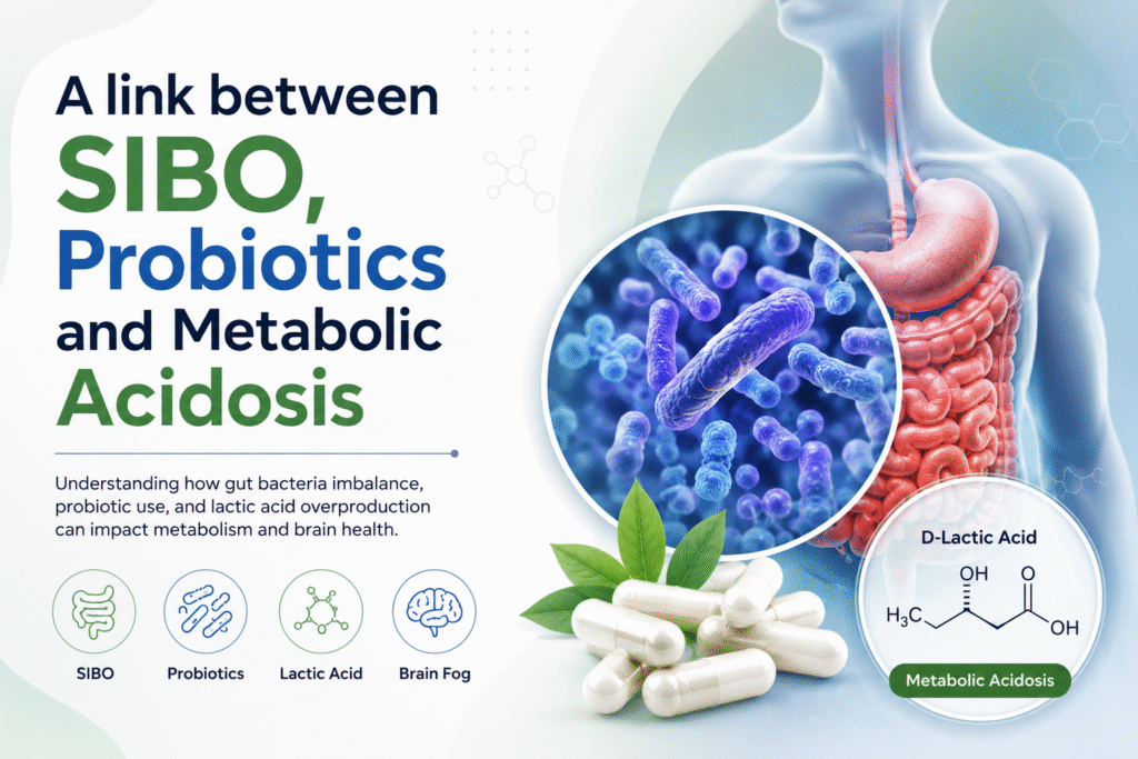
A link between SIBO, Probiotics and Metabolic Acidosis Lactic acid is typically produced in excess at a rate of about 20 mmol/kg per day and enters the bloodstream, where it is primarily metabolized by the liver and kidneys. While some tissues can use lactate as a substrate and oxidize it to carbon dioxide (CO2) and water, only the liver and kidneys possess the necessary enzymes for gluconeogenesis from lactate. The tissues that typically produce excess lactic acid include the skin, red blood cells, brain tissue, muscles, and the gastrointestinal tract. During intense exercise, skeletal muscles are the primary source of elevated circulating lactate, which usually returns to normal levels if hepatic metabolism is unimpaired. Elevated lactate levels may result from increased production, decreased clearance, or a combination of both (2). SIBO and D-Lactic Acidosis D-lactic acidosis is an underrecognized complication that can occur in patients with small intestinal bacterial overgrowth, or SIBO, and dysbiosis of the gut microbiome (1). Abdominal bloating, gas and distension are common gastrointestinal symptoms that are caused by many conditions including carbohydrate intolerance and small intestinal bacterial overgrowth (SIBO) (3). Brain Fogginess (BF) describes a constellation of symptoms comprised of mental confusion, impaired judgment, poor short-term memory, and difficulty with concentration, which is often transient and disabling. Previously, similar symptoms, along with slurred speech and gait disturbances have been described in patients with short bowel syndrome (4,5). These patients were found to have metabolic acidosis with elevated levels of D-lactic acid in the serum. The L-isomer of lactic acid is the main form of lactic acid in the human body and is produced by the enzymatic (L-lactate dehydrogenase) reduction of pyruvate. D-lactate is produced in much smaller quantities by the enzyme D-2-hydroxyl acid dehydrogenase that also metabolizes D-lactate in the human liver (4).  In a healthy gut, D-lactate levels remain low because production is limited and clearance is efficient. However, in SIBO or severe dysbiosis, there is an overgrowth of bacteria—particularly species like Lactobacillus—that ferment carbohydrates into D-lactate (2). In short bowel syndrome, if the colon is colonized by D-lactate producing bacteria, the delivery of large amounts of unabsorbed carbohydrate causes rapid fermentation, gaseous distension, and production of large amounts of d-lactic acid overwhelming hepatic clearance (6) and causing D-lactic acidosis and encephalopathy. When excess carbohydrates reach the small intestine, these bacteria rapidly convert them into D-lactate, which is then absorbed into the bloodstream. Unlike L-lactate, the human body metabolizes D-lactate more slowly, leading to accumulation. This buildup can result in D-lactic acidosis, characterized by metabolic acidosis and a range of neurological symptoms. Patients may present with confusion, brain fog, slurred speech, ataxia, or unusual behavior—often episodic and sometimes mistaken for psychiatric or neurologic disorders. Risk factors include short bowel syndrome, but it is increasingly recognized in patients with SIBO and significant gut dysbiosis, especially when high carbohydrate intake fuels bacterial fermentation. Probiotics and D-Lactic Acidosis The enzyme lactate dehydrogenase catalyzes the interconversion of pyruvate and lactate, stereospecific for the L-isomer (L-lactate), the predominant form synthesized and metabolized in humans. Although D-lactate is not a major component of normal mammalian metabolism, it can be produced in significant amounts by certain bacteria and yeast, particularly within the gastrointestinal tract. Probiotics are considered to be safe and beneficial including improvement in gut barrier function and gut transit (7). Although a meta-analysis of 57 studies indicated that probiotics are safe (8), caution against its use has been recommended in subjects who are immunosuppressed, pregnant, and with structural heart lesions, acute abdomen, neutropenia, chemotherapy and radiotherapy (8,9).  Recently, probiotic use has been implicated in the production of D-lactic acidosis, both in short bowel syndrome patients and in the first 2 weeks of life in infants who were fed probiotic-containing formula (10,11). Typically, D-lactic acidosis is caused by the fermentation of ingested carbohydrates by D-lactic producing bacteria such as lactobacillus and bifidobacterium in the bowel (4,5).  Lactobacillus species and bifidobacterium are the most common bacteria in probiotic formulations (7,12) and are felt to be useful in the treatment of irritable bowel syndrome, inflammatory bowel disease, and other intestinal problems (13). Both bacteria produce D-lactic acid. D-lactic acidosis has been described with Salmonella enteritidis and with probiotics (14). Probiotics are designed to deliver bacteria to the colon but whether this is achieved has not been reliably shown (7,12-15). In contrast, they may colonize the small bowel, especially in the presence of dysmotility or low acid conditions that favor bacterial overgrowth.  Interestingly, some Lactobacillus species such as Lactobacillus GG only produce L-lactate (14,16). Hence, both D-lactate and L-lactate should be measured when assessing this condition, and the most practical way for its diagnosis is to administer a carbohydrate meal and assess D-lactate in urine and L-lactate in blood, over the next 3 h along with breath samples for hydrogen and methane. Management focuses on reducing D-lactate production and correcting the underlying imbalance. This may include dietary carbohydrate restriction, targeted antibiotics for SIBO, probiotics in select cases, and correction of acid-base disturbances. Reference Study  Rao et al. (1) studied a cohort of patients with an intact gut who reported brain fogginess associated with unexplained abdominal bloating, pain, gas and distension, and in whom there was evidence of probiotic use, D-lactic acidosis and SIBO. They found that over 2/3rd of patients with brain fogginess exhibited D-lactic acidosis, along with significantly higher prevalence of SIBO when compared to those without brain fogginess. Symptoms improved with the use of antibiotics and stopping probiotics. Hence, they advise caution against excessive and indiscriminate use of probiotics especially without a well-defined medical indication, and particularly in patients with gastrointestinal dysmotility, and/or those using long term PPI and opioids (1). Conclusion In summary, D-lactic acidosis is a clinically important but often overlooked consequence of SIBO and gut dysbiosis.  D-lactic acidosis is a rare but clinically significant subtype of lactic acidosis, typically observed in patients with short bowel syndrome or other forms of gastrointestinal malabsorption. In these individuals, undigested glucose and starch are fermented by colonic bacteria into various organic acids, including D-lactic acid—an isomer that humans metabolize poorly. Systemic absorption of

Scroll to Top