Author name: Vishi Sethi

Blog

Can SIBO Predispose you to Nutrient Deficiencies?

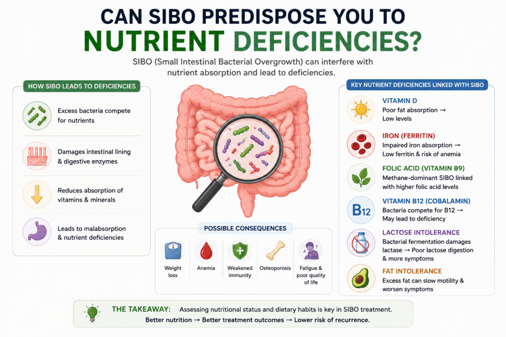
Can SIBO Predispose you to Nutrient Deficiencies? Small intestinal bacterial overgrowth (SIBO) is a pathology of the small intestine and may predispose individuals to various nutritional deficiencies. The studies suggest that the subtypes of SIBO (Hydrogen and Methane dominant) may have varying effects on dietary intake, leading to a range of biochemical deficiencies. Conversely, specific dietary patterns (such as dairy, fiber, gluten, fat etc.) may predispose one to the development of a SIBO subtype.  Diet is a modifiable factor that plays a crucial role in shaping the composition, diversity and stability of the gut microbiota (1). A diet rich in fiber and plant-based foods, supplemented with prebiotics and low in choline and fat is generally acknowledged to predispose one to a healthy microbiota (3). Conversely, a regimen deficient in fiber, fermentable oligosaccharides, disaccharides, monosaccharides and polyols (FODMAPs), and characterized by an elevated intake of omega-6 fatty acids typical of a Western dietary pattern, may predispose one to dysbiotic conditions (3,4). However, an approach in which proper care for gut microbiota nutrition is neglected may lead to nutritional deficiencies and microbiota alteration (5,6,7). Moreover, these issues can result in malabsorption, leading to nutritional deficiencies (vitamin A, D, E, B12), hypoproteinemia, anemia and weight loss (8,9,10). While SIBO is not a life-threatening condition, it can worsen the patient’s health and underlying comorbidities (11,12,13). Individuals experiencing gastrointestinal symptoms frequently turn to prolonged dietary restrictions, sometimes neglecting the significance of restoring microbial equilibrium in the gut (14,15). Furthermore, patients blindly choose to consume only permitted products, often leading to an inadequate intake of fiber, calcium or iron due to poor meal planning (16,17,18,19).  However, it remains unclear whether a specific subtype of SIBO may predispose patients to a particular clinical presentation (i.e., nausea, diarrhea, constipation), which influences their dietary choices and patterns, or whether a patient’s baseline diet and nutritional intake may increase the risk of developing a specific SIBO subtype. A study by Wielgosz et al. (1) investigated the possible correlations between dietary intake (protein, fat and carbohydrates), serum levels of biochemical parameters (vitamin A, D, E, B12, folic acid, calcium, ferritin and iron) and SIBO subtypes in newly diagnosed SIBO patients. Adult participants between the ages of 18 to 65 years old presenting with existing abdominal symptoms occurring at least three times per month in the past six months were recruited. The study only included those individuals who newly had SIBO, confirmed through a non-invasive hydrogen–methane breath test with lactulose substrate (LHMB) and had not yet started antibiotic therapy or the use of the often-recommended elimination diet (1). The results are as follows: The H+/M+ group – was associated with low serum vitamin D (p < 0.001), low serum ferritin (p = 0.001) and low fiber intake (p = 0.001).  The M+ group – was correlated with high serum folic acid (p = 0.002) and low intakes of fiber (p = 0.001) and lactose (p = 0.002). More than 50% of M+ dominant patients displayed a tendency towards iron deficiency. The H+ group – was associated with low lactose intake (p = 0.027). None of the H+-dominant patients exhibited iron deficiency. Moreover, over 50% of the H+ group exhibited optimal ferritin levels. Furthermore, it was noted that higher concentrations of exhaled H2 were associated with significantly lower levels of serum vitamin D. Lower serum ferritin concentrations were associated with higher levels of H2 gas production (p = 0.001, r = −0.5648). And it was observed that higher folate levels were solely associated with increased CH4 concentrations (p = 0.002, r = 0.6367). Regarding the intake of macronutrients and selected vitamins and minerals, four correlations were identified by the same study (1). In the H+/M+ group and M+ group, an association was observed between fiber intake and exhaled CH4 gas. The higher the concentration of CH4 gas production, the lower the intake of fiber in both groups. Lactose intake correlated with both H2 and CH4 gas production. In the H+ group, higher levels of exhaled H2 gas were associated with lower lactose intake in the diet (p = 0.027, r = −0.6338). Conversely, in the M+ group, a lower lactose intake was correlated with higher levels of exhaled CH4 gas (p = 0.002, r = −0.6444). The remaining parameters, including energy intake, protein, fat and carbohydrate intake, as well as vitamins A, D, E, B12, folate, and minerals such as calcium and iron, were not correlated with exhaled H2 or CH4 (1) Core-relation of Biochemical Parameters with SIBO Subtypes: Vitamin D – In the proximal part of the small intestine, SIBO may instigate the deconjugation of bile acids which reduce the solubilization of dietary fat within micelles (20). This alteration leads to the presence of unconjugated bile acids and a shift in their site of reabsorption, consequently leading to reduced absorption of fat-soluble vitamins (A, D, E) (20). Wielgosz et al. (1), in their research found that in the H+/M+ group, the concentration was 7155 ppm/min AUC, compared to 6160 ppm/min AUC in the H+ group and 2306 ppm/min AUC in the M+ group. However, the values only reached a statistically significant difference in the H+/M+ group and M+ group. It is important to consider that methane-producing Archaea are a hydrogen cross-feeder; hence, the lack of differences in H2 breath concentrations between the H+/M+ and H+ groups may be attributed to this phenomenon (21). Although each of the patients examined was recently diagnosed with SIBO, evidently, higher H2 levels in exhaled breath contributed to the impairment of vitamin D absorption, and a more pronounced state of dysbiosis could potentially affect this group (1). However, to date, few studies have assessed vitamin D levels in patients with SIBO, and none of these studies has correlated vitamin D levels and SIBO subtype. In a study conducted by Zhang et al., significantly lower serum vitamin D levels were observed in pregnant patients with gestational diabetes and SIBO, compared to a control group without SIBO (23). Among patients with concurrent SIBO and chronic pancreatitis or systemic sclerosis, no differences were noted in vitamin D levels compared to a group without SIBO (24,25)  One consequence of vitamin D deficiency is hypocalcemia,
Blog

Can Hypothyroidism predispose you to SIBO?

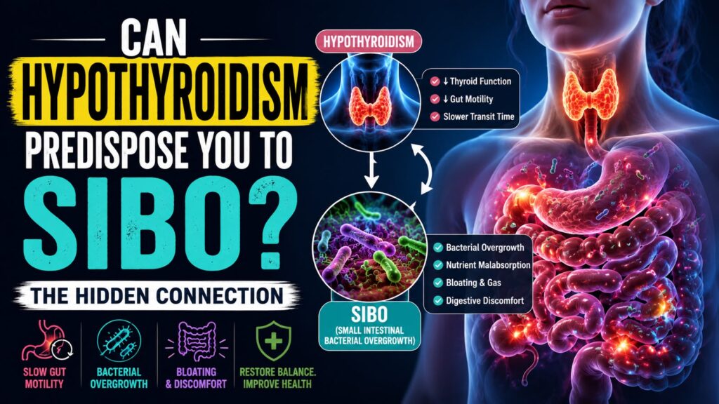
Can Hypothyroidism predispose you to SIBO? How Hypothyroidism Contributes to SIBO? (SIBO in Hypothyroidism) It has been reported that SIBO may be present in more than half of the patients with hypothyroidism (5). Recent studies suggest that small intestinal bacterial overgrowth (SIBO), a disorder of excessive bacteria in the small bowel, may also be associated with hypothyroidism (6-8). Some studies have suggested that hypothyroidism results in decreased gut motility, which increases the risk for the development of SIBO (1,3). The objective of this article is to understand the association between hypothyroidism, altered GI motility and development of SIBO. Small intestinal bacterial overgrowth (SIBO) is defined as an abnormally high bacterial population level in the small intestine. SIBO occurs when mechanisms controlling enteric bacterial growth are disturbed. In SIBO, bacteria such as Gram-negative coliforms overgrow in the small intestine, leading to gastrointestinal symptoms such as abdominal pain, bloating, and gas (12,13). One of the most common mechanisms that lead to SIBO is the small intestinal dysmotility. Thus, small intestinal dysmotility constitutes one of the risk factors for development of SIBO (9,10,11). Other risk factors include failure of the gastric acid barrier, anatomic alterations or impaired immunity (10). On the other hand, Hypothyroidism is one of the most common endocrine conditions globally (14-16). Hypothyroidism can affect multiple organ systems, resulting in symptoms associated with decreased quality of life, including fatigue, weakness, memory issues, hair loss, hypersensitivity to cold, dry skin, and depression (17,18).  Hypothyroidism is known to affect the entire gastrointestinal system and cause hypomotility (19-21). A reduction in the motor activity of stomach, small intestine, and colon has been reported in previous studies. It has been found that hypothyroid patients show significant reduction in gastric emptying and thus have a delayed intestinal transit time (19-22). Changes in the motor activity of the digestive system may result in gastric or abdominal distension and constipation in hypothyroidism (20,23). Clinically, constipation and abdominal distension are known to be the typical gastrointestinal (GI) symptoms of SIBO as well. Thus, intestinal motor dysfunction associated with hypothyroidism could predispose you to bacterial overgrowth. Study references: A study done by Olga et al. reported that Hypothyroidism prominently reduces esophageal and gastric motor activity and can cause gastrointestinal dysfunction. The study group included 30 females with primary hypothyroidism and 10 healthy females. They used – Gastroesophageal scintigraphy method, which is a noninvasive, simple, physiological method to use for evaluating esophagogastric motility and can be helpful in selecting treatment regimens for hypothyroidism (3). Another study by Wei et al. evaluated the incidence and risks of subsequently developing SIBO, and changes in small bowel microbial populations, in subjects with hypothyroidism or autoimmune thyroiditis (2). The findings from this study suggest that there is an increased risk of subsequent development of SIBO in individuals with hypothyroidism, and that this risk is increased in individuals with autoimmune thyroiditis. Moreover, the overgrowth of specific Gram-negative coliforms may be involved, as greater prevalences of Klebsiella species and lesser prevalence of Escherichia/Shigella are seen in the core duodenal microbiome of individuals with hypothyroidism and SIBO as compared to individuals with SIBO alone (2). How Gut Dysbiosis contribute to Hypothyroidism? (Hypothyroidism in SIBO) Recent studies indicate that gut microbial dysbiosis may influence the onset and progression of hypothyroidism through mechanisms involving immune dysregulation, increased intestinal permeability, chronic low-grade inflammation, and impaired nutrient absorption. Also, microbial metabolites such as short-chain fatty acids and bile acids are increasingly recognized as modulators of thyroid hormone metabolism, tissue sensitivity, and enterohepatic circulation (4). Additionally, Microbiota influence the uptake of minerals relevant to the thyroid gland including iodine, selenium, zinc, and iron. All of them play a role in supporting thyroid function and there is a clear link between thyroid dysfunction and altered levels of these minerals (4). Iodine, iron, and copper are crucial for thyroid hormone synthesis, selenium and zinc are needed for converting T4 to T3, and vitamin D assists in regulating the immune response. Those micronutrients are often found to be deficient in autoimmune thyroid diseases (AITDs), resulting in malfunctioning of the thyroid. (4) The inflammatory regulation of an impaired microbiota and their negative influence on the immune system seems to be likely to promote autoimmune diseases such as autoimmune thyroid diseases (AITD) (24). Hashimoto’s thyroiditis (HT) is the most common thyroid disorder worldwide with a general prevalence of around 10–12% and is characterized by chronic inflammation, leading to hypothyroidism, and often, destruction of the thyroid gland (25,26). Graves’ disease (GD) is another AITD which has a prevalence of 1–1.5%. It has been proposed that the interaction of genetic predisposition, immune impairment, and environmental factors (i.e. micronutrients, gut microbiota) play an important role in the pathogenesis of these diseases. (4) Furthermore, intestinal bacteria play a key role in vitamin synthesis (such as vitamin K, folic acid, vitamin B2, B3, B5, B6, B7, and B12 (27,28) digestion of dietary fibers, regulation of the immune response, and mental disorders (4). Heme iron Fe2+, is an important source for both the human and the intestinal microbiota. Studies have reported that iron is essential for bacterial growth and iron availability influences the composition of the microbiota because some bacteria have developed better heme-catching mechanisms. On the other hand, microbiota can increase iron bioavailability in the colon through lowering of pH via the production of short chain fatty acids (4). Some Bacteria can make siderophores (such as enterobactin) which are high-affinity proteins for iron that acquire Fe3+ (dietary non-heme iron), especially in iron-poor environments. These are chelators that pull iron away from insoluble compounds and move it into a form that bacteria can take up. Humans developed a defense protein called lipocalin-2 to sequester siderophores and limit microbial growth (24,29,30).  Pathogenic strains particularly grow well in heme-rich conditions, due to their efficient heme capturing ability (29). Many enteric gram-negative bacteria, including Salmonella, Shigella, and pathogenic E. coli require iron for their virulence and colonization (31,32). On the other hand, Beneficial commensal gut bacteria from genera Lactobacillus and Bifidobacterium require less or no iron (33). In mice, Constante et al. demonstrated that a heme-rich diet decreases microbial diversity

Blog

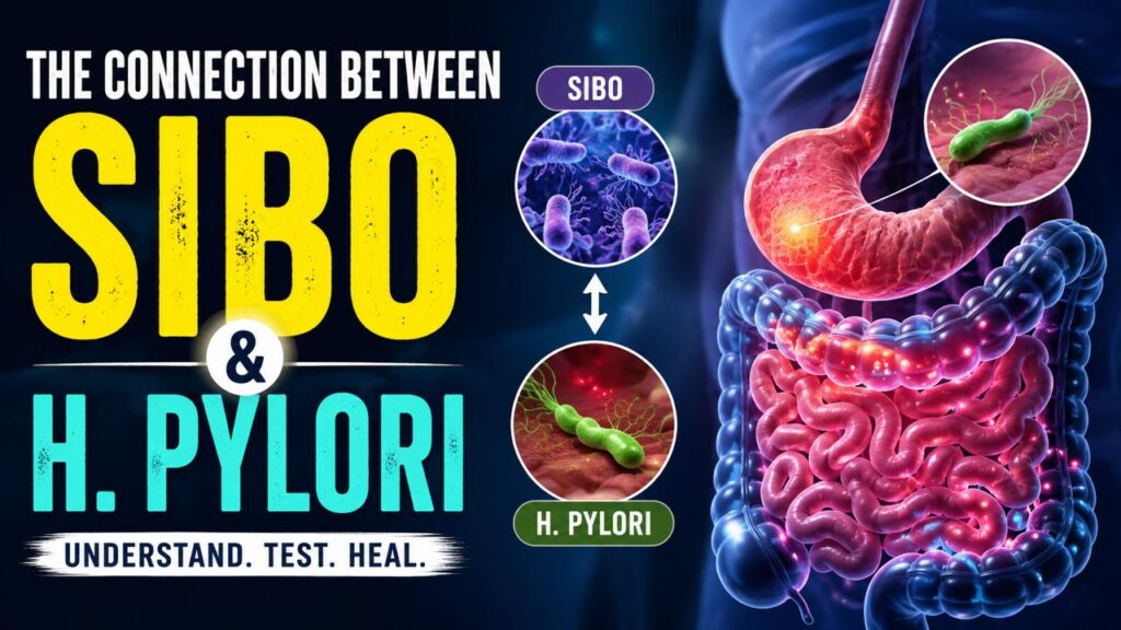
The connection between SIBO and H. Pylori

The connection between SIBO and H. Pylori Helicobacter pylori (H. pylori) infection is very common and affects a significant proportion of the world population with a prevalence rate in the United States between 20% and 40% (1). In contrast, the prevalence of small intestinal bacterial overgrowth (SIBO) in the general population is not well understood. There can be a coexistence of both disease states in a given patient, and their clinical symptoms may also overlap with one and another. However, there are no clear clinical guidelines for testing and treating SIBO in patients with H. pylori infection. Therefore, this article explores – the relationship between H. Pylori and SIBO as well as their association with other pathologies. The Big Question – If H. Pylori infection is associated with SIBO? Can H. Pylori infection predispose you to SIBO? Does H. Pylori infection create an environment ripe for SIBO? H. pylori enters the body through the mouth, moves through the digestive system, and infects the stomach or the first part of the small intestine. The spiral-shaped bacterium uses its tail-like flagella to move around and burrow into the stomach lining. This damages the gut lining and causes inflammation. H. pylori is a nasty pathogen that can persist in the stomach of infected persons for a lifetime, if left untreated. It provokes a chronic gastric inflammatory response, resulting in the development of several gastric pathological conditions including superficial gastritis, chronic atrophic gastritis, peptic ulcers, gastric cancer, and mucosa-associated lymphoid tissue (MALT) lymphoma (44). Although the majority of individuals colonized by H pyloriare asymptomatic, a proportion of patients develop peptic ulcers (duodenal and gastric), and an even smaller proportion develop gastric cancer. Globally, H pylori is the major cause of gastric cancer and has been classified as a Class I carcinogen by the WHO. Epidemiological studies have revealed that people with lower vegetable and micronutrient intake may be at increased risk of H. pylori infection (44).  To successfully colonize the host and establish infection, H. pylori must be able to withstand an acidic stomach and adhere to host cells. In order, to survive the harsh acidic environment of the stomach, it produces a substance called Urease, a cytoplasmic enzyme. This substance helps them neutralize the stomach acid and is largely responsible for H. pylori’s acid tolerance (1). During active H. Pylori infection, this gram-negative bacteria uses urease to hydrolyze (convert) urea into ammonia and carbonic acid/carbon dioxide in the stomach. The ammonia byproduct buffers gastric acid leading to an increase in stomach pH to protect the organism and allow further proliferation (1). Over time, atrophy of the gastric mucosa (thinning, shrinking or wearing of gut lining) occurs permitting further multiplication of the bacteria. The preferred treatment of H. pylori infection includes a course of proton pump inhibitor (PPI) therapy (which further raises gastric pH) (5) and antibiotic agents which are also known to cause dysbiosis (because they wipe out good and bad bacteria both) and consequent gastrointestinal symptoms (6,7). Both mucosal atrophy and gastric pH alterations have been proposed to predispose patients to SIBO (1). In the general population, gastric secretions are strongly acidic with a pH range of 1 to 2. In non-H. pylori infection individuals, daily administration of 20 mg omeprazole (PPI) has been shown to increase gastric pH by 2 to a pH range of 3 to 4 (1).  During H. pylori infection, individuals receiving this same dose of omeprazole showed increased stomach pH by a total of 4 to a pH range of 5 to 6 (8). Within the pH range of 5 to 6, enteric bacterial load can increase by as much as 1000-fold (11). This causes abnormalities in the intestinal flora. H. pylori often infects the stomach at a young age and significantly reduces the post-infection Firmicutes to Bacteroidetes ratio at the phylum level (9). Successful eradication of H. pylori increases the amount of Bifidobacterium in the intestinal flora (10).  These bacteria (H. Pylori) are predominantly gram-negative anaerobes that produce gas with the fermentation of carbohydrates (when you eat food as in carbs). This gas fermentation allows for the detection of H. pylori infection by the urea breath test and the detection of SIBO by the hydrogen breath test (1). With that said, both bacterial load and the gas they produce contribute to the nonspecific constellation of gastrointestinal complaints (such as bloating, indigestion and acidity) as described in SIBO and H. pylori infection.  A meta-analysis study conducted by Liao L et al. (2) pooled the results of eight eligible observational studies and found that H. Pylori infection may be related to SIBO in adults. Hence, the detection of SIBO should be considered for patients with digestive symptoms and HP infection. The study (2) results suggest that HP infection is associated with a higher prevalence of SIBO in younger adults (mean age <48 years) as compared to older adults (mean age >48 years). Subgroup analyses further indicated that the association was not significantly affected by the country of study, comorbidities, exposure to proton pump inhibitors, or methods of evaluating HP infection and SIBO (2). Helicobacter pylori infection is also a risk factor for various gastric diseases, such as chronic gastritis, peptic ulcers (gastric and duodenal), atrophic gastritis, and gastric cancer (12,13). Increasing studies suggest that besides gastric diseases, H. Pylori infection may also be involved in the pathogenesis of some intestinal disorders (14). For example, H. Pylori infection has been linked to the risk of colorectal adenomas (15) and colorectal cancer (16), as well as functional disorders such as IBS (17), suggesting a close relationship between H. Pylori infection and disturbed intestinal homeostasis. According to research studies, impaired gastric motility and/or acidity will likely boost bacterial growth in the small intestine and increase colonization (9,10) thus, creating an environment ripe for SIBO.  Numerous studies have suggested an association of SIBO with, altered anatomy, hypochlorhydria (low stomach acid), dysmotility, immune deficiencies, and small intestinal disease and PPI use (18,19). Physically, the small intestine has relatively low-level colonized bacteria compared to the colon (8). Recent evidence from preclinical and clinical studies suggests that excessive bacterial growth in the small intestine, which is called small intestinal bacterial overgrowth (SIBO) (20),

Blog

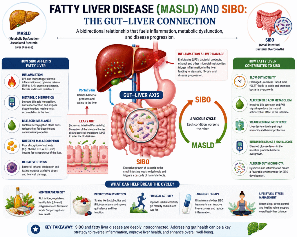
EXPLORING THE LINK BETWEEN FATTY LIVER AND SIBO

Fatty Liver Disease, MASLD, and SIBO Fatty liver disease, particularly Nonalcoholic Fatty Liver Disease (NAFLD), now known as Metabolic Dysfunction-Associated Steatotic Liver Disease (MASLD), has become a major global health concern. In recent years, MASLD has been the second leading cause of end-stage liver disease worldwide (28). Increasing evidence reveals a fascinating connection between NAFLD and gut health, specifically a condition known as Small Intestinal Bacterial Overgrowth (SIBO). The correlation between small intestinal bacterial overgrowth (SIBO) and nonalcoholic fatty liver disease (NAFLD) has gained heightened acknowledgment, especially in the late phases of liver disease. Today, we explore the scientific links between these two conditions and why understanding this relationship matters. Understanding The Gut-Liver Axis The gut epithelium is a natural barrier that selects entry of useful substances present in the lumen, as nutrients, and keeps at bay bacteria, their bio-products and other potentially harmful elements. Tight junctions, specialized intercellular structures, assist this control. Derangement of the homeostasis between bacteria and the host, as occurs in SIBO (enhanced amount and/or changes in the type of bacteria in the gastrointestinal tract), may cause disruption of the intercellular tight junctions and subsequent increase in intestinal permeability leading to bacterial translocation (BT), i.e., transportation of bacteria and bacterial products from the intestinal lumen into the blood (4). The portal vein and the hepatic artery supply blood to the liver. The portal blood contains products of digestion and microbial products derived from the gut microbiota. This blood is carried to the liver. Therefore, Liver is the first site of exposure and filtration that consists of microbial products from the gut, such as LPS, lipopeptides, unmethylated DNA, and double-stranded RNA, which may evoke inflammatory reaction contributing to the progression of the liver disorder (4). This bidirectional relationship of the gut ecosystem and liver is imperative both physiologically and pathologically. Generally, the liver receives rich nutrients, microbial metabolites, and subproducts from the intestine and secretes bile into the small intestine (5). An integrated gut barrier also protects against toxins to maintain internal homeostasis. This gut-liver axis is regulated and stabilized by a complex network of metabolic, immune, and neurosecretory interactions between the gut, microbiota, and liver. Disruption of this equilibrium may lead to gut dysbiosis and liver injury (5). SIBO as we know is the clinical manifestation of gut microbial dysbiosis. Therefore, the bidirectional relationship between Small Intestinal bacterial overgrowth (SIBO) and fatty liver disease, particularly non-alcoholic fatty liver disease (NAFLD), is characterized by mutual influences through gut-liver axis dysfunction, inflammation, and metabolic disturbances. Association between SIBO and Fatty Liver Small intestinal bacterial overgrowth (SIBO) is a condition marked by excessive growth of microbes in the small intestine, resulting in various digestive issues including bloating, satiety, and malabsorption. In healthy people, the small bowel has a relatively low bacterial concentration, around 103–104 colony-forming units per milliliter (CFU/mL) (1,6). However, when this balance is disrupted, bacteria from the colon or oral cavity can colonize the small intestine, resulting in SIBO. Factors contributing to this condition include reduced gastric acid production, impaired intestinal motility, insufficient production of bile and dysfunction of the ileocecal valve (1,7). It may present in a range of symptoms, from moderate pain to severe nutritional deficiencies, weight loss, and shortages in crucial minerals and vitamins, including vit B12, A, D, E, iron, choline, calcium, fats, carbohydrates, protein and bile salt deconjugation (1,8). However, it has been shown that intestinal dysbiosis, endotoxemia (bacterial toxins in blood) and bacterial translocation may contribute to inflammation and Insulin Resistance (3,9,10,11,12). This directly seems to disrupt the functioning of the gut–liver axis, which may influence the incidence and progression of NAFLD (3,13). Non-alcoholic fatty liver Disease is the most frequent cause of chronic liver sickness globally, with a spectrum spanning from simple steatosis to inflammation of the hepatocytes, fibrosis, cirrhosis, and even hepatocellular carcinoma (1,14). The link between SIBO and nonalcoholic fatty liver disease (NAFLD) has attracted increased attention since studies show that the gut-liver axis plays a significant role in the pathophysiology of steatosis liver disease (1,15). The transfer of bacterial metabolites from the stomach to the liver may promote scarring and inflammation, thereby aggravating liver damage (1,16). How SIBO affects Fatty Liver?   Inflammation – SIBO leads to an overgrowth of bacteria in the small intestine, causing increased intestinal permeability “known as leaky gut or gut barrier dysfunction.” This allows bacterial endotoxins, especially lipopolysaccharides (LPS), to enter the bloodstream and reach the liver via the portal vein, triggering chronic liver inflammation. These endotoxins and bacterial products activate inflammatory pathways (immune responses) in the liver, releasing proinflammatory cytokines (e.g. TNF-α, IL-6) which promote fat accumulation, insulin resistance, liver inflammation, fibrosis, and disease progression in NAFLD. Example – It has been demonstrated in animal models that a four-week HFD (high fat diet) increases LPS contained in the gut microbiota and plasma LPS concentrations two to three times, which is considered metabolic endotoxemia. The induction of metabolic endotoxemia in mice, by continuous subcutaneous infusion of LPS for four weeks, was followed by a rise in the following parameters: fasting glycemia, insulinemia, markers of inflammation, liver triglyceride content, liver insulin resistance, and whole body, liver and adipose tissue weight gain in a similar amount as occurred in HFD fed mice (4,17). Large amount of fructose consumption is also related to increase in endotoxin serum levels, proinflammatory response and steatosis. It was demonstrated in an elegant study conducted by Bergheim et al. (4,18) that mice fed with fructose showed increased endotoxin levels in the portal blood, and higher intrahepatic lipid accumulation, lipid peroxidation and TNF-α expression. Metabolic disruption – SIBO also disrupts bile acid metabolism, nutrient absorption, and adipose tissue function, contributing further to liver fat deposition and dysfunction. Bile acids – Bile acids are synthesized in the liver from cholesterol, conjugated (joined) with glycine or taurine, and secreted into the small intestine, where they help digest fats and maintain gut microbial balance through their antimicrobial properties (3). In a healthy gut, most bile acids are absorbed in the ileum, while the

Blog

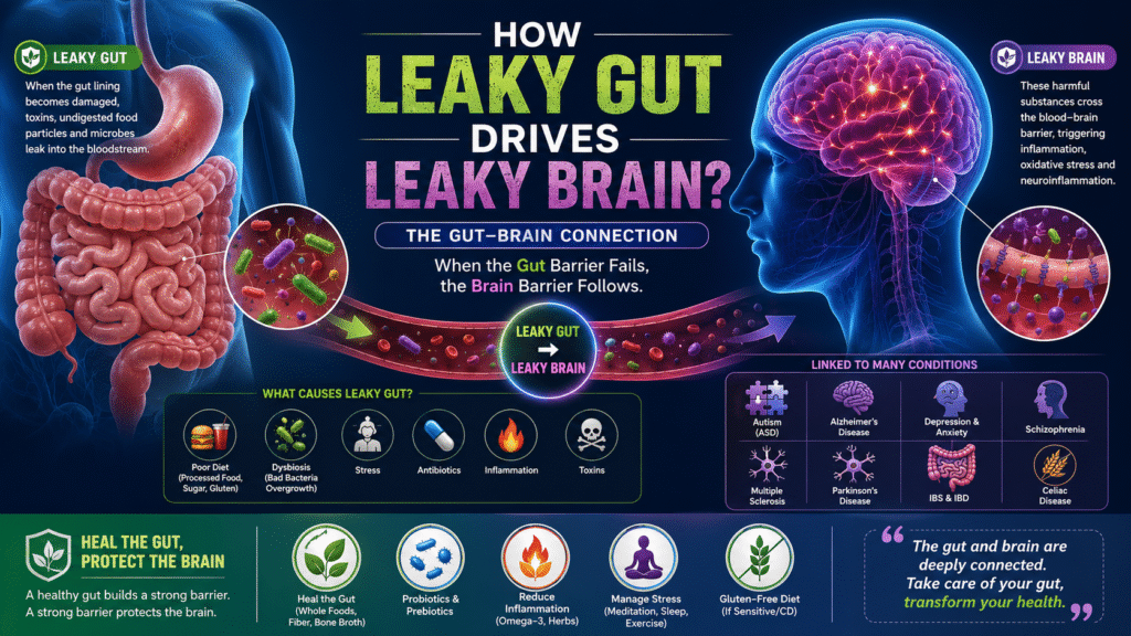
How Leaky Gut Drives Leaky Brain?

How Leaky Gut Drives Leaky Brain? This article focuses on the possible neuropsychological basis of leaky gut; leaky brain disease; and the microbiota’s contribution to inflammation, gastrointestinal, and blood-brain barrier (BBB) integrity.  As already indicated, in my other article titled as “leaky gut syndrome,” various diseases have been related to dysbiosis of the intestinal microbiota, microbial translocation, and dysfunction of the intestine’s barrier function. Among them, we can highlight IBS, IBD, Obesity, Chronic Heart Failure, Autism, Alzheimer’s Disease, Cancer, Diabetes, and Autoimmune Diseases like Type 1 Diabetes and Celiac Disease (1). Critical to any discussion on leaky barrier systems are pathogens, which, unlike commensals, have evolved elaborated mechanisms to target host barrier integrity and disseminate systemically to invade deeper tissues and organs. This pathogen gains entry into the blood stream by acting through type IV pili on bacterial surfaces, interacting with molecules on endothelial cells to disrupt the tight junctions and occasionally escape the mucosal barrier to enter the bloodstream and pass into the meninges of the brain and its surrounding membranes to cause disease and breach the blood–brain barrier (BBB) (1,4). To understand a possible connection between leaky gut and possible leaky brain, let’s examine the barriers involved in physiological conditions.  Vital organs and biologic systems have developed barriers to host’s tissues from infection. The notable barriers are the blood brain barrier (BBB), gastrointestinal blood barrier (GBB), blood ocular and blood retinal barriers, blood placenta and blood testis barriers, the blood thymus barrier, and the blood–lung or airway barrier. Each of these barriers protects vulnerable and sensitive organs and systems (1). A key component for the brain is the neurovasculature, which limits blood brain barrier (BBB) permeability and prevents transport of large molecules, many small molecules, and bacteria from entering the brain. Inflammation disrupts BBB and appears to be central to brain and blood brain barrier (BBB) involvement (1).  Many diseases and physiological stressors that affect the Central Nervous System (CNS) also alter the functional integrity of the BBB (9,10). They affect the barrier’s ability to selectively restrict passage of substances from the blood to the brain. To add to this, hypoxia (lack of oxygen supply) and/or inflammation and inflammatory process alter the permeability properties and contribute to the pathophysiology of CNS diseases, leading to altered delivery of therapeutic agents to the brain (5). Selective permeability is important and accomplished through tight junctions, composed of endothelial cells and smaller subunits anchored into the endothelium together with transmembrane proteins, such as junctional adhesion molecule, occludins, adherens, and claudins, for example. The junctional proteins in the brain are like those of the small intestine (1). Tight junctions help protect the brain from toxins, chemicals, and pathogens that might be circulating in the bloodstream. Together with selective transport proteins, the barriers allow nutrients, oxygen, amino acids, some drugs, and glucose to enter the cerebrospinal fluid and prevent hydrophobic molecules from passing into the interfaces of blood–cerebrospinal fluid barriers, namely CSF and choroid plexus. At the same time, it allows the diffusion of many small polar molecules, dissolved gasses, hormones, and hydrophilic molecules (1). In the gut, the barrier between the body and a lumenal environment is formed by gastrointestinal mucosa, buffering nutrients, microorganisms, and toxins. The barriers are semipermeable, thus allowing efficient transport of nutrients across the epithelium, while excluding entry of potentially harmful small molecules and organisms. The exclusionary properties of the gastric and intestinal mucosa are referred to as the gastrointestinal blood barrier (GBB) (1,6). As the barriers share common proteins and features, there is no doubt they may be susceptible to similar mechanisms of compromise or breach, either biochemically or physically. This fact underlies one basis for a plausible LEAKY GUT LEAKY BRAIN SYNDROME. A functional blood–brain barrier is essential to maintaining central nervous system (CNS) homeostasis. BBB weakening may be a result of a disturbance in the endothelial cells due to P-glycoprotein dysfunction (7). If toxins or microorganisms breach the epithelium, they have unrestricted access to the systemic circulation. In the brain, this can occur with disruption of endothelial cells and astrocytes and involve inflammation (8). In the gut, the alimentary canal is lined by epithelial cells that form the mucosa and, with few exceptions, the gastrointestinal epithelium is tied contiguously through tight junctions, where diversity among epithelial cells affect specific barrier functions.  When the Gut-Blood-Barrier (GBB) is breached, there are differences in localization of bacterial species. When a breach occurs, commensal bacteria deposit in the lymphatics and are not found in the blood stream, but species like Salmonella, which by definition are pathogenic – can establish infections in the blood, liver, or other organs. We have evolved with commensals and not pathogens through colonization resistance and other mechanisms, which is one reason why commensal bacteria end up in the nearby lymph nodes and are not found in distal organs despite being proximal to the epithelial linings. Other stressors, such as glycoxidative stress (AGEs), diabetes, prolonged hyperglycemia, and obesity, are risk factors for gut–blood barrier disruption. To resolve these processes, restoration of epithelium (repair of gut lining) must happen, which can be rapid and is accomplished by a process called restitution. Advanced glycation end-products (AGEs) and crosslinking in diabetic complications and with aging may also be a mechanism for barrier protein damage with advanced age (11) and could be mediated through glycotoxins from food. Google definition of Glycotoxins – are compounds, most commonly Advanced Glycation End-products (AGEs), that form when sugars react with proteins or fats. High levels of glycotoxins are linked to oxidative stress, inflammation, and chronic diseases like diabetes, heart disease, and Alzheimer’s. Brain Disorders and Gut Microbiota A dysfunction of the blood brain barrier leading to a ‘leaky brain’ can be linked to various neurological diseases, including autistic spectrum disorder (ASD) (12), dementia, Alzheimer’s disease, depression, and schizophrenia (20,13)  A breakdown in the blood brain barrier was observed in patients with major psychiatric illnesses (14) indicating that the blood–brain barrier may become ‘leaky’ in select neurological diseases that have an immunologic component, such as multiple sclerosis (MS) (15,16),

Blog

Biofilm Formation and Gastrointestinal Disorders

As per the reports of the National Institute of Health (NIH), about 80% of human infections affecting the gastrointestinal, genitourinary (UTIs), respiratory systems, oral mucosa and teeth, eyes, middle ear and skin are caused due to BIOFILM FORMATION by biofilm-associated microorganisms. The ability to form biofilms is a universal attribute of bacteria, and biofilms play a role in several infections including – infection of indwelling medical devices, wound infections, bacterial carditis (heart-infection), otitis media (middle ear infection), dental carries, and lung infections of cystic fibrosis patients (13). Gut microbiota dysbiosis, mucus disruption, and epithelial invasion are associated with pathogenic biofilms that have been linked to gastrointestinal disorders such as irritable bowel syndrome (IBS), Inflammatory Bowel Diseases (IBD), gastric cancer, and colorectal cancer (2). Intestinal biofilms are highly prevalent in ulcerative colitis and irritable bowel syndrome (IBS) patients, and most endoscopists have observed such biofilms during colonoscopy, in the gastrointestinal environment (GI tract) (2). IBS and IBD are the two most frequent GI disorders, together affecting >10% of the Western population. A recent clinical study revealed endoscopically visible mucosal biofilms in 57% of IBS, 34% of ulcerative colitis (UC), and 22% of Crohn’s disease (CD) patients (6% healthy; 976 patients in Austrian cohort and 450 in German cohort) (2,12). Biofilm-positive UC and IBS patients had an altered microbiome compared with biofilm-negative individuals, a finding independent of disease state (2.12). Antibiotics and food additives might contribute to the reduced microbial diversity and biofilm formation, and it is plausible that food industrialization is linked to biofilm formation frequency, aligning with the higher prevalence of IBS and IBD observed in Western populations (2). What are Gastrointestinal Biofilms? Gastrointestinal biofilms are matrix-enclosed, highly heterogenic and spatially organized polymicrobial communities that can cover large areas in the gastrointestinal tract. The human gastrointestinal (GI) tract is the alimentary canal extending from the mouth to the anus and is the most densely inhabited environment of the human body. The GI tract harbors a profusion of microorganisms with different lifestyles called the gut microbiota, accounting for around 30% of the human microbiome. Along the GI tract, bacterial density increases, with the highest density in the colon (109–1011 bacteria/mL). The interplay of bacteria with the environment and the host affects the microbiota’s phenotypical occurrence and composition. Many gut microbes live as free-floating cells in the lumen, whereas others adapt higher-ordered structures termed biofilms (2,3,4,5) (as shown in the picture). This picture illustrates – How Bacteria adopt different lifestyles in their natural habitats from single planktonic cells to biofilm communities. (Ref 2) The mucus layer predominantly comprises dynamic mucin glycoprotein sheets coating the epithelial surface, forming the main barrier between the intestinal epithelium and luminal content (2,6). It effectively protects the host from digestive enzymes, acids, microbial by-products, food-associated toxins, pathogens, and microbial infiltration, preventing infection and inflammation (2,6,7). A compromised mucus layer or defects in mucus production can facilitate bacterial colonization and mucosal biofilm formation (2,6,8). Biofilm formation on the outer mucus layer can lead to mucosal invasion and bring bacteria close to the epithelium, an event that is disease-associated (9,10). Polymicrobial biofilms naturally grow throughout the gastrointestinal tract, both at the epithelial surface and in the lumen as mucin-attached and food particle-attached colonies. In simple words – A biofilm is a complex multi-cultural community of microorganisms, such as bacteria, fungi, or algae, that stick to each other and often adhere to a surface within a slimy, self-produced matrix known as the extracellular polymeric substance (EPS) or “slime.” This matrix, made up of sugars, proteins, lipids, and DNA, protects the microbes and helps them survive in harsh conditions by providing a shield against antibiotics, disinfectants, and the host immune system. How does Biofilm Formation Happen? Let’s liken the multispecies bacterial biofilm to a city – where bacteria settle selectively, limit settlements of new bacteria, store energy in exopolysaccharide, and transfer genetic material horizontally all for the good of the many (1). There are several steps that we must take to optimize our lives in a city. The first is to choose the city in which we will live, then we must select the neighborhood in the city that best suits our needs, and finally we must make our home amongst the homes of many others. Occasionally, when life in the city sours, we leave. The same steps occur in the formation of a bacterial biofilm as shown in the picture below (1). To give a larger picture – Free floating Bacteria (known as planktonic cell) attach to a mucosal surface, form microbial colonies, flagellin is reduced and create a hydro-gel like structure called exopolysaccharide or EPS matrix to protect themselves from host’s immune system and antibiotics. This EPS layer secretes extra polymeric cellular substances such as water, polysaccharides, lipids, proteins, and extracellular DNA resulting in a three-dimensional network, known as EPS matrix, that provides mechanical and chemical stability. The matrix protects against host defense mechanisms, mechanical forces of intestinal peristalsis, and antimicrobials through slow or incomplete drug penetration (1). Formation of a Bacterial Biofilm (Picture taken form reference 1) The Life Cycle and Architecture of Gastrointestinal Biofilms include the following steps and mechanism: The Biofilm Life cycle – (Picture taken from Ref 2) Picture below illustrates the appearance of GI biofilm inside the colon and its matrix composition (Ref 2) A. Gut bacteria and biofilm appearance – Bacterial communities are distributed throughout the digestive tract and adapt distinct phenotypes (planktonic, biofilm, and biofilm-dispersed). Most of these communities are free-floating (planktonic state) but also occur as mucosal biofilms or as aggregated biofilms to food particles and mucins. Bacteria from mucosal biofilms can invade the host mucus layer and bring them in close contact with the epithelium, a state that is often associated with a reduced host immune and antimicrobial response and the onset of disease (2). B. Biofilm matrix composition – The biofilm matrix predominantly comprises water and biopolymers, including polysaccharides, proteins, lipids, and extracellular DNA, forming a hydrogel-like structure. Bacterial cells are embedded in this matrix and together form

Blog

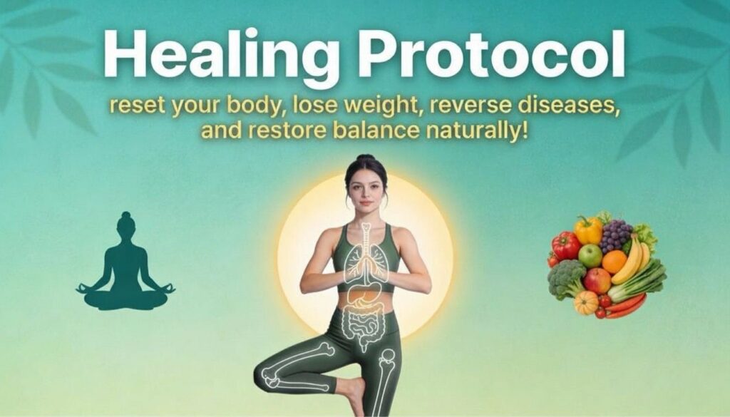
Healing Protocol to reset your body, lose weight, reverse diseases, and restore balance naturally and powerfully!

Healing Protocol to reset your body, lose weight, reverse diseases, and restore balance naturally and powerfully! Weight gain isn’t your fault – it’s your body’s SOS signal for help! In a world overflowing with quick fixes and fad diets, true healing often begins when we shift our focus from restriction to restoration.  The body isn’t your enemy – it’s a miraculous system designed to repair itself when given the right environment. This means –  “You don’t need a stricter diet – you need a deeper healing protocol!” A healing protocol isn’t about counting calories or chasing the scale; it’s about identifying and reversing the root imbalances that caused weight gain and disease in the first place.  By nourishing your liver, balancing your gut microbiome, calming inflammation, and realigning your hormones through clean foods, mindful movement, and deep rest, you can trigger your body’s innate ability to heal.  This isn’t about temporary change – it’s about reversing disease and dropping weight by – fixing what’s really broken inside! Let’s dive deep into “the targeted Healing Protocol” that can help you lose weight, detoxify fatty liver, reset metabolism, reverse chronic conditions and metabolic disorders (like Cholesterol, BP, Type 2 diabetes, Thyroid, Obesity etc.) and naturally guide your body back to its ideal weight – one mindful step at a time. Follow the steps below to – reclaim your health, energy, and peace from the inside out. Let’s talk about “WHAT TO EAT, WHEN TO EAT AND HOW TO EAT?”  Step 1 – Upon arising in the morning (let’s say you wake up at 7 am) drink one glass of Lukewarm water.  Reason – this water does not get absorbed in the body rather it flushes out toxins and cleanses your kidneys Step 2 – Do some form of exercise – for atleast one hour (from 7 to 8am) preferably – meditation, yoga and pranayama. Reason – yoga and pranayama help in reducing stress. Stress as we know is a major underlying factor in the onset and progression of many diseases – for it has a direct impact on your digestion and can mess up your gut.  Step 3 – After getting done with the exercise at 8 am – Drink a cup of Milk Thistle Tea.  Reason – Milk Thistle is a potent ayurvedic herb that detoxifies your liver and removes toxins from your body. Step 4 – Sit in the sun for 15 to 20 mins. You can also sit in the sun while sipping milk thistle tea.  Reason – Sitting in the sun nourishes both mind and body. It enhances digestion and sets the mood and tone for the day. It also promotes Vit D synthesis, strong immunity and detoxication through mild sweating.  Step 5 – After drinking milk thistle tea, one hour later at 9 am, have a probiotic drink. Probiotics add good healthy bacteria to your gut. Which aids in digestion, strengthens the lining of your gut, decreases inflammation, and improves insulin sensitivity.  Here are my top 3 recommendations for the probiotic drinks –  Homemade kanji – made from beetroot and carrots Kefir – can be coconut kefir, goat’s milk kefir or kefir water Buttermilk – made from plain homemade yogurt or coconut yogurt. I suggest you add a spoon of flax seed powder, or chia seeds (soaked overnight) or psyllium husk fiber to your probiotic drink. All these fibers act as prebiotic (fodder) for the good bacteria in your gut.  Step 6 – One hour later at 10 am, have one fruit or a small plate of salad with few nuts and seeds. Make sure you eat different types of fruits and salad with different types of nuts and seeds for at least 7 days in a week and then repeat. This will help in diversifying your microbiome.  Note – you can choose to eat just salad at this hour and eat a fruit after lunch in the evening before 4pm. This is because some salads don’t pair well with fruits and so fruit should be eaten alone with nuts and seeds. Also, since salad is must for it adds fiber to the diet, it should be eaten as an appetizer at least one hour before lunch.  Make sure the nuts and seeds are soaked overnight before consumption.  For example – On  Mon – eat – 1 apple or cucumber and carrot salad with 5 to 7 almonds Tues – eat – 1 banana with a spoon of sunflower seeds Wed – eat – 1 pear – with a spoon of pumpkin seeds Thurs – eat – a bowl of papaya – with 5 cashews and so on Reason – eating fruits and nuts once a day is must for fruits are light to digest, high in water content, and rich in fiber and micronutrients.  Nuts and seeds are a great source of omega 3 fatty acids, protein and vitamin E – which are healthy for your brain and heart.  Step 7 – Have an early lunch between 11 to 11:30am. Let’s call it a brunch.  Now the Lunch should be Nutri-dense! Your plate must contain –  A bowl of vegetables (any two seasonal vegetables mixed) – green leafy vegetables like – kale, arugula, broccoli, cauliflower, spinach, zucchini, squash, pumpkin, water cress, asparagus, celery, peas, carrots, bell-peppers, Brussel sprouts, artichoke, sweet potatoes, mushrooms, okra, bottle guard/ridge gourd/Indian round guard (aka Ghiya/Tinde/Tori), bitter gourd, Potatoes and taro root (aka Arbi) in moderation (no more than once per week), onions and tomatoes in moderation (cooked only) A bowl of legumes – consume all lentils basically – black eyed peas, chana dal, moong dal, moong chilka dal, arhar or toor dal, kidney beans, pinto beans, garbanzo beans.  A bowl of protein – such as tofu, edamame, soya, eggs, red -meat in moderation, chicken, turkey, fish, chick-peas, homemade paneer or cottage cheese, quinoa etc. as per your digestion preference.  Whole grains – now if you have a weak gut – I suggest you eat gluten free grains like – millets,

Blog

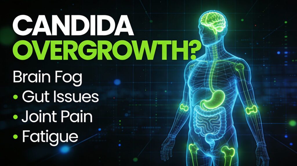
DO YOU HAVE CANDIDA OVERGROWTH?

DO YOU HAVE CANDIDA OVERGROWTH? If you are experiencing chronic symptoms ranging from Brain Fog, Intestinal Discomfort, Joint Pain to Fatigue, but your doctor is at a loss to explain the reason, Candida Overgrowth could be to Blame!  What is Candida? Humans host a complex microbial ecosystem composed of bacteria, fungi, viruses, protists and archaea – each playing key and interconnected roles in maintaining health and supporting biological functions. The gut microbiome varies greatly between individuals, both in size (total microbial load) and composition (species diversity), due to differences in physiology, diet, lifestyle, and environment.  Candida is a yeast-like fungus that naturally lives on and inside the human body – primarily in the mouth, gut, skin and vaginal tract – without causing harm under normal circumstances (1). However, Candida can become opportunistic under certain conditions and cause infection – when the normal balance within the microbiome is disrupted or the host’s immune system is weakened (1). The main drivers of candida overgrowth include poor diet, antibiotics, stressful lifestyle, alcohol, high blood sugar, contraceptive hormones, immune suppression and gut disrupting drugs.  Over 150 Candida species have been recognized to date. But Candida Albicans is the most frequently associated with HUMAN INFECTIONS.  It is a dimorphic Gram-positive yeast, not acid-resistant, saprophyte (fungus that lives on dead or decaying matter) in 40–75% of healthy humans (28, 29). And is responsible for over 80% of human yeast infections (28,30). Among humans – Candida Albicans is the most studied and clinically relevant – an overgrowth of which affects millions of people; many of whom are unaware of the cause of their symptoms. In fact, it is estimated that one in three people suffer from yeast overgrowth due to Candida Albicans! Candida albicans is a common opportunistic yeast that causes oral thrush, vaginal infections, and gut issues! The genus name Candida is attributed to the traditional “white robes” worn by Roman candidates (candidatus) running for public office. The term albicans is derived from another Latin word albico/albicatus, which means “to be white” or “verge on white.” In essence, the term Candida albicans is redundant meaning “white to be white” and is associated with white color [2]. What is Candida Overgrowth or Pathogenicity? Candida overgrowth happens when the natural balance between Candida yeast and beneficial bacteria in the body is disturbed, allowing candida to multiply excessively and cause infection. This can occur in various parts of the body such as the gut, mouth, throat, or genital area.  When Candida overgrows, it disrupts the normal microbial balance and can lead to symptoms and infections collectively known as Candidiasis.  In most individuals with a healthy immune system, C. albicans is a harmless microbe that exists in harmony with other members of the gut flora that keep candida under control. Candida begins to overgrow when the environment in the body changes due to the following factors: Repeated use of broad-spectrum antibiotics (kills both good and bad bacteria creating room for fungus to grow) Changes in the host immune system (e.g., during stress, poor diet and lifestyle, infection by another microbe, or immunosuppressant therapy) Variations in the gut environment (e.g., shifts in gut pH, gastritis, or nutritional deficiencies) and, Conditions such as diabetes, HIV/AIDS, chemotherapy, organ transplantation Damaged physical barriers (damaged mucosal barriers such as mouth, gut, genital surfaces due to trauma, chemotherapy, or medical devices for example catheters) These conditions favor the trigger of fungal proliferation causing Candida to grow out of control, invade deeper tissues and turn into an            aggressive pathogen causing wide range of infections (1). These infections range from: superficial mucosal and dermal infections, such as oral thrush, diaper rash or skin rash, and vaginal yeast infections (75% of women will have yeast infection or UTIs at least once in their lifetime),  to more serious blood/organs related and deep tissue infections with sizable mortality rates (approaching 47% in some cases) (1). While women are more susceptible to vaginal or genital yeast infections (also known as UTIs in lay term), men and kids can also be infected.    Certain factors, such as prolonged antibiotic use, increase the risk of candida overgrowth for both men and women. Prolonged Antibiotics use promotes yeast (fungal) infections, including gastrointestinal (GI) Candida overgrowth and penetration of the GI mucosa making the host more susceptible to yeast infections. Candida albicans may also play a role in the persistence or worsening of some chronic inflammatory bowel diseases (IBD) (5,6). Many of the illnesses and symptoms that plague men and women and children today – from fatigue, bloating, and weight gain to prostates, brain fog, arthritis, allergies, ear infections and depression can be traced back to surprising factor – an overgrowth of yeast called Candida Albicans (5,6). In addition to these concerns, candida has also been linked to arguably more serious and debilitating illnesses such as autoimmune diseases like multiple sclerosis, fibromyalgia, autism, rheumatoid arthritis, lupus and mental illness (5).    Infection caused by Candida are especially serious in immunocompromised individuals (such as those with diabetes, nutrient deficiencies, AIDS/HIV or those undergoing anticancer chemotherapy/radiation immunosuppression therapies) and healthy people with implanted medical devices such as catheters, pacemakers, prosthetics, heart valves, dentures etc. (1,6). Candida’s ability to shift from a harmless commensal to an aggressive pathogen exemplifies its opportunistic nature – it takes advantage of weak immune system or disrupted microbial imbalance to cause a disease.    Factors that lead to Candida Overgrowth    Diet high in sugar and refined carbs – Candida thrives on sugar.  Repeated Antibiotic use – Antibiotics kill beneficial and non-beneficial bacteria (not fungi), that normally keep candida in check. This disrupts the microbial balance allowing the yeast to overgrow.  Low stomach acid (Impaired Upper Digestion) – impacts gut microbiome and again makes the environment favorable for candida overgrowth Weakened Immune System and Health conditions – Conditions such as HIV/AIDs, diabetes, cancer, chemotherapy, and use of immunosuppressive drugs increase susceptibility to candida Excess alcohol consumption – Excess alcohol can lead to leaky gut, change gut flora and thus act as a fuel source for candida overgrowth. Estrogen Dominance – Changes

Blog

How did I beat Mold (Ochratoxin A)?

How did I beat Mold (Ochratoxin A)? My Mold Toxicity Story! All of this started when I was 16 years old (back in year 2000), studying in a boarding school (India) and was severely Constipated. To treat my Constipation, Doctors put me on Laxatives (Yellow tablets by Homeopathy, Cremalax and Dulcolax by Allopathy) and my health started deteriorating day by day. My symptoms were – Loose/watery stools, Low energy, Sinusitis, Brain Fog, Feeling Lethargic, Chronic Fatigue, Breathing problem, Forgetfulness, Problems in reading Textbook, Irritation on silly things, Bone and Muscle weakness, Dry Cough, Unexplainable Weight Gain, Intensified Sugar Cravings, Brain feeling deprived of nutrition even if I ate 5 meals a day, Problem in waking up early, Mood Swings, Feeling Frustrated, Inflammation in the joints and gut, Binge eating and Emotional eating too, Heartburn, Gas/Acidity/GERD. I SERIOUSLY NEEDED HELP!! After going to numerous Allopathic, Ayurvedic and Naturopathic doctors for 18 years in vain – I finally decided to see a Functional Medicine Doctor at the age of 37 (I moved to America in 2009 at the age of 25). I had seen a few functional medicine doctors earlier but they could not help much. They could not diagnose what was going on with me exactly. Finally, after a lot of research, I found two great Functional Medicine (FM) Doctors that had all the answers for me. First FM Doctor diagnosed me with SIBO (Small Intestinal bacterial overgrowth (Methane and Hydrogen dominant), Leaky gut issues, deficiency of trace minerals, imbalance in gut flora, hiatal hernia and gastritis. Second FM doctor diagnosed me with SIFO (Small intestinal fungal overgrowth) also known as Candida overgrowth and Liver and Gallbladder Congestion. Now I knew I was in the right hands! I hope you all know that SIBO is a medical condition in which – bacteria starts to populate in your small intestine where all of your nutrients are absorbed – leading to chronic malabsorption of nutrients from food. The right place for the bacteria is to reside in the large intestine. Same goes with SIFO – which refers to small intestinal fungal overgrowth which is also very common. Now, the first FM doctor began a treatment for SIBO. Her approach was – biofilm disruption for two weeks with LOW FODMAP DIET followed by antibiotics for two weeks. But did it work? NOPE it didn’t! Why didn’t it work? I followed her protocol exactly as she recommended:     1. Took Interface plus – a biofilm disruptor for two weeks   2. Ate LOW FODMAP food during those two weeks – I had turned vegan by that time so it was kind of hard because that made my food choices more restrictive. So all I ate was tofu and black gram tortilla with some LOW FODMAP vegetables. I used to rotate between salads and vegetables. 3. Then in the third week I started my course of antibiotics (Rifaximin and Metronidazole) for two weeks. I saw the difference within first week. I lost some weight and was passing lot of gas which of course was a good sign of those bad bugs being killed. Didn’t put any sugar in my mouth. 4. However, in the second week of antibiotics, I started craving sugar. So I made myself some orange juice at home thinking it’s a LOW FODMAP food. BUT THAT TURNED OUT TO BE THE BIGGEST MISTAKE! 5. My SIBO relapsed immediately – flaring up a reaction instantly. I knew I had messed up. Sugar is what the bad bugs love. Also, another  mistake I made was I didn’t add a binder and didn’t do enema daily. However, I was doing all other spiritual practices like – meditation, yoga and breath work since SIBO is supposed to be a cause of Stress and Fear. (I will talk more about on the spiritual aspect of SIBO and Candida in my next blog post) 6. Anyways, finally completed the course for two weeks with no long lasting results. Discussed with my FM doctor, told her I can’t eat tofu and the same vegetables anymore. My body had given up. My kidneys and liver were rejecting protein completely. Also, I had this complaint that whenever I ate sugar, especially, it came in my eyes like it made my eyes blurry. It also caused lot of congestion in my chest area underneath my rib cage specifically. 7. She just thought they were all signs of bloating due to SIBO. So, she came up with a new plan. She decided to put me on an elemental diet  for two weeks. It’s a liquid diet which contains pre-digested nutrients which do not reach your small intestine so bad bugs never get to feed on them. The idea is to starve the bad bugs in the intestine and kill them. 8. I had a great experience with elemental diet. Lost 7 kgs of weight, gave rest to my liver and kidney, felt amazing in brain, had lot more  energy and my concentration improved. But my chest pain was still there. It just won’t go away even with this diet. Another major side effect  I had of this diet was infection in my ears. However, I ignored it thinking we will deal with this later. Let’s just focus on beating SIBO first. 9. In the second week of my elemental diet program, I accidentally put a piece of almond in my mouth and there it was again, the reaction  flared up. I was like “you kidding me – it came back with just a piece of almond?” Told my doctor and she said it’s hard to do it. I was            disappointed with this response. I knew there was something more going on in my body besides SIBO. 10. Well this time she tweaked the plan again and she put me on Herbal antimicrobials (Candibactin AR and BR) for 4 weeks. I started taking those coz they are easy on your stomach and liver. But also started doing my own research

Blog

Panchakarma Treatment

Panchakarma Treatment Feeling sluggish, depressed, unmotivated? Having difficulty with emotional issues, digestion, or concentration? You may benefit dramatically from panchakarma: detoxifying through Ayurveda. Panchakarma (translated as five cleansing actions in Sanskrit) is a gentle, yet profound purification therapy, designed to reduce the body of toxins, which cause disease and discomfort. Feeling good already? Panchakarma is supreme for disease prevention and life extension and is the heart of healing with Ayurvedic medicine. When the energetic forces known as vata, pitta, and kapha get out of balance, it lowers our digestive fire, and toxins are created. The ama enters the blood stream, clogging the channels, and lodges into joints, muscles, organs, and reproductive tissue, etc. This is detrimental to our prana (life-force or energy), tejas (metabolic energy), and ojas (immunity). When using Ayurveda to heal, we can choose palliative care—which includes diet, meditation, yoga, pranayama, herbs, etc.—and/or take a deeper path of cleansing. Cleansing enables us to get to the root of imbalances—rather than reducing symptoms—and can heal chronic conditions. For thousands of years, Ayurveda has emphasized that the stress created by our minds directly affects our Gastrointestinal Tract, causing inflammation and slow digestion, ultimately creating ama. The ama causes us to hold on to emotionally driven habits that ultimately lead to disease and imbalance. Detoxing with panchakarma allows us to unravel these patterns, increase our energy andmental clarity, and move forward with our dharma and our dreams. Panchakarma resets digestion and allows the body to do what it’s supposed to—detox naturally. The complete process of Panchakarma involves the following three steps:    Poorva Karma This is a preparatory procedure required before the main treatment, to soften the tissues so that the lipid-soluble toxins deposited into them are liquefied and flow back into the digestive tract. From here, they can be eliminated. This treatment prepares the patient mentally and physically for the main procedure of Panchakarma. It involves three procedures: Pachan Karma – improves digestion with herbs and fasting so that the patient can digest the ghee (clarified butter) which is provided to liquefy the fat-soluble toxins. Snehan Karma – medicated ghee is given to the patient in increasing doses to aggravate and liquefy the fat-soluble toxins deposited in the deep tissues. Swedan Karma – a full body steam bath is given to the patient thereby opening the body channels and allowing the heat to liquefy the toxins further. This facilitates their movement from the tissues to the digestive tract. Pradhan Karma This is the Panchakarma, a five-step procedure which is highly individualized depending on the needs, age, digestive strength, immune system, and other factors. This intense Panchakarma procedure can only be done under the guidance of an Ayurvedic practitioner. The five karmas to cleanse the complete body are:    Vamanam (therapeutic emesis) – induced vomiting which helps clear the upper GI tract to the duodenum (end of the stomach) and a part of the respiratory tract.  Virechanam (purgation) – induced purgation clears the GI tract from the duodenum to the exit. Anuvasana (enema using medicated oil) – the oil enema helps lubricate the rectal area and remove the lipid soluble waste out through the anus. Nasyam – nasal inhalation of medicated substances which help clear the respiratory tract and the para nasal sinuses. Paschaat Karma This is a post-therapy dietary regime to restore the body’s digestive and absorptive capacity to its normal state. It includes rejuvenating treatments, lifestyle management, diet management, and intake of herbal supplements. It includes the following procedures: Sansarjan Karma – food therapy after detoxification, which aims at gradually increasing the patient’s diet from liquids to semi-solids to a normal diet. Rasayan Adi Prayogam – a rejuvenating rasayan therapy which aids in increasing natural immunity and enhancing your general well-being. Shaman Chikitsa – a pacification therapy with herbs and lifestyle management. It is important to note that a panchakarma procedure is designed specifically for an individual after a thorough physical examination and pulse diagnosis.

Scroll to Top