July 28, 2026

Blog

LEAKY GUT SYNDROME –

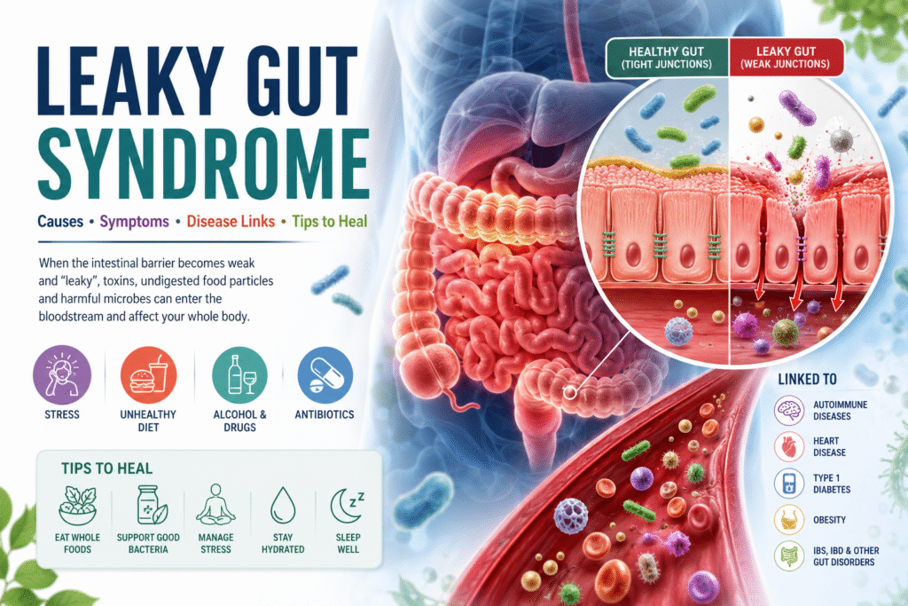
LEAKY GUT SYNDROME – Causes, Symptoms, Disease links and Tips to Heal The human body is in daily contact with potentially toxic and infectious substances in the gastrointestinal tract (GIT). The GIT protects the intestinal integrity by allowing the passage of beneficial agents and blocking the path of harmful substances.  Under normal conditions, a healthy intestinal barrier (gut lining) prevents toxic elements from entering the blood stream. However, factors such as stress, an unhealthy diet, excessive alcohol, antibiotics, and drug consumption can disturb the composition of the intestinal microbiota (gut flora) and homeostasis of the intestinal barrier of the intestine, leading to increased intestinal permeability.  The Intestinal Hy-permeability can allow the entry of harmful agents through the junctions of the intestinal epithelium, to leak into the blood stream and affect various organs and systems and is known as LEAKY GUT SYNDROME (LGS)! An increase in intestinal permeability is a sign of a disturbed intestinal barrier (2). According to the leaky gut syndrome (LGS) hypothesis, intestinal hyperpermeability may allow the entry of harmful microorganisms, toxins, or undigested food particles through the junctions of the intestinal epithelium, reaching the bloodstream and being able to affect the hormonal, immune, nervous, respiratory or reproductive systems (3). Thus, dysfunction of the intestinal epithelial barrier and increased permeability results in a “leaky gut” that is associated with intestinal disorders such as inflammatory bowel disease (IBD), irritable bowel syndrome (IBS), alcoholic liver disease (ALD), nonalcoholic fatty liver disease (NAFLD), steatohepatitis, liver cirrhosis, and collagen diseases (1). Leaky gut syndrome is also associated with extra-intestinal diseases (diseases that are not related to intestinal disorders) such as heart diseases, obesity, type 1 diabetes mellitus, and celiac disease (1).   Thus, the mucosal barrier is crucial to protect the body from exogenous harmful biological and chemical agents, such as microorganisms and environmental pollutants. The function of gastrointestinal epithelial barrier is to protect against the entry of foreign antigens and microorganisms, while allowing the absorption of essential nutrients, water and electrolytes (1,57).  The intestinal barrier consists of four components: microbial barrier, biochemical barrier, physical barrier and immune barrier (1,57). The microbial barrier is the intestinal microbiota, located in the lumen of an intestine. The microbiota produces many metabolically active compounds that show antimicrobial activity and affect the function of the entire intestinal barrier. Commensal bacteria digest certain food components and also compete with pathogens for nutrients (1,57). The biochemical barrier is mucus, which contains about 98% water (1) and, among others, mucins, glycoproteins, IgA antibodies, antimicrobial substances, produced by microorganisms—bacteria, viruses, fungi and intestinal cells. Mucus coats epithelial cells and protects them from the harmful effects of pathogenic microorganisms and toxic substances (57).  The physical (epithelial) barrier is an essential component of the entire intestinal barrier. It consists of a single layer of specialized cells: enterocytes, goblet cells (produce mucins), Paneth cells (produce antimicrobial peptides and proteins), enteroendocrine cells, M cells and intestinal stem cells. These cells undergo renewal every 3–5 days. Epithelial cells have a variety of functions and are closely interconnected (1,57).  The immune barrier is associated with the presence of lymphoid tissue in the intestines known as gut-associated lymphoid tissue (GALT). The GALT system is located in the mucosa and submucosa of the intestines, directly beneath the epithelial cells. This system consists of intraepithelial lymphocytes (IELs), Peyer’s patches, which are clustered lymphoid papules, and lymphocyte clusters. The GALT system has also been found to contain antigen-presenting cells (APCs), T lymphocytes, B lymphocytes, plasma cells, as well as macrophages, mast cells and dendritic cells (DC). The secretory IgA antibody (sIgA) is synthesized in the intestine in particular (1,57). This picture is taken from reference 1 This picture is taken from reference 57 The intestinal barrier is a selective barrier—its function is to allow the transport of digested food essential for the body’s function, but at the same time to keep harmful substances and microorganisms in the intestinal lumen, which requires strict regulation of the barrier’s permeability (1,57). This transport is regulated by tight junctions (TJ) (1,57). The TJ between enterocytes play a key role in providing an intestinal barrier. These junctions are composed of proteins, including occludin, claudin and junctional adhesion molecules (JAMs) and peripheral proteins known as zonula occludens (ZO-1, ZO-2, ZO-3), which bind to actin filaments (1). The primary role of zonulin is to dynamically open and close tight junctions between epithelial cells, thereby regulating paracellular permeability. When zonulin is released and its pathway is activated, it triggers intracellular signaling (including protein kinase C and cytoskeleton rearrangement) that leads to reversible disassembly of tight junction proteins such as ZO‑1 and occludin. Increased zonulin production has been observed under the presence of certain bacteria and food components, such as gliadin peptides found in gluten. Excessive release of zonulin results in weakening of TJ and consequent passage of antigens into the vicinity of immune cells and into the circulatory system. As a result, local inflammation develops and activated immune cells and cytokines can affect other organs or trigger immune-related diseases, e.g., autoimmunity (58). Elevated zonulin levels and increased permeability have been associated with several chronic inflammatory and autoimmune conditions, including celiac disease, type 1 diabetes, inflammatory bowel disease, and some neuroinflammatory and neurodegenerative disorders (59). Because of its strong association with barrier dysfunction, zonulin is being explored as a biomarker of impaired gut barrier function in various autoimmune and chronic inflammatory diseases. Pollution and climate change, chemical compounds commonly used in industry and households, ecosystem changes, unhealthy diet, and stimulants, mainly alcohol, tobacco and e-cigarettes, may disrupt the epithelial barriers of the skin and mucosal surfaces. Air, water and food pollution, microplastic particles, nanoparticles, household chemicals and tobacco smoke are the most common epithelial barrier disrupting factors (57). Therefore, pathogenesis of inflammatory bowel and leaky gut diseases is associated with multifactorial causes as discussed below.  Gut Microbiome and Leaky Gut The gut has more than 100 trillion bacteria (4), with an aggregate biomass of approximately 1.5 kg (5) composed of more than 200 microbial strains in an individual and
Blog

Exploring the Link Between Candida Overgrowth and Fatty Liver Disease

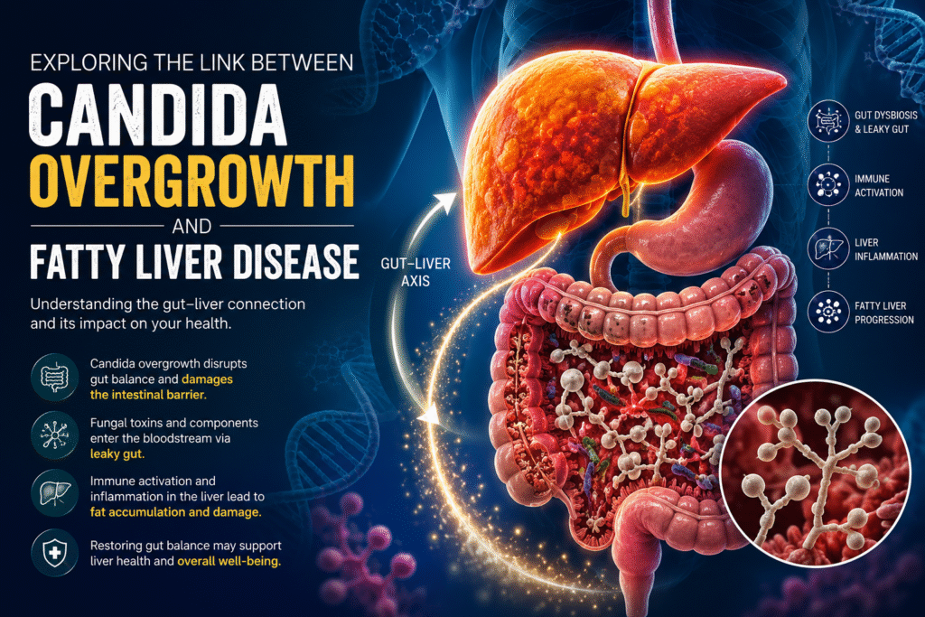
Exploring the Link Between Candida Overgrowth and Fatty Liver Disease What is a Fatty Liver Disease? Fatty liver disease, particularly Nonalcoholic Fatty Liver Disease (NAFLD), is a widespread metabolic condition impacting millions worldwide. Emerging research highlights not only the role of bacteria but also fungi, especially Candida species, in influencing liver health. This blog explores how Candida overgrowth may be linked to fatty liver and what it means for your health. Note – NAFLD is now called MASLD (Metabolic Dysfunction Associated Steatotic Liver Disease) as it is linked to metabolic issues like obesity, diabetes, and high cholesterol, characterized by fat buildup in the liver. What is Candida Overgrowth? Candida is a type of yeast that naturally lives in the gut, mouth, and other parts of the body. Under normal conditions, it coexists peacefully with the body’s microbial community. However, when Candida grows excessively—a condition known as Candida overgrowth—it can disrupt gut balance, potentially leading to digestive issues, immune dysfunction, and increased intestinal permeability (leaky gut). Interactions between intestinal fungal community (aka. mycobiome) and liver are anatomically and functionally bidirectional as explained below: Antigens derived from the gut commensal fungi can cross the gastrointestinal barrier and be transported via the portal vein to the liver, thus having an impact on its function. The immune cells in the liver can contribute to the host homeostasis through immune responses to the intestinal fungi (1,2). Thus, it has become apparent that gut mycobiome has a profound influence in modulating local as well as peripheral immune responses. However, like bacteria, fungi can be beneficial to host immunity. But they may also have deleterious effects (1,3) when the host immune system is compromised, or repeated rounds of antibiotics are used (4). This causes changes in the composition of the intestinal microbiota, promoting a series of liver diseases through the “entero-hepatic axis.”  Candida’s role in Fatty Liver Pathology Candida overgrowth is linked to fatty liver disease – alcoholic fatty liver disease -ALD (5) and non-alcoholic fatty liver disease – NAFLD (1) through mechanisms involving inflammation and immune response triggered by fungal components. A study (5) observed that alcohol-dependent patients displayed reduced intestinal fungal diversity and Candida overgrowth. Compared with healthy individuals and patients with non–alcohol-related cirrhosis, alcoholic cirrhosis patients had increased systemic exposure and immune response to mycobiota (1). Candida albicans and other fungi are found enriched in the gut microbiota of people with NAFLD, especially those with inflammation and advanced fibrosis (1). Inflammation is known to play a major role in the progression of NAFLD and NASH. Liver immune cells when exposed to fungal antigens and fungi derived metabolites elicit anti-inflammatory cytokines and chemokines, some of which can lead to liver damage (1,2). The fungal cell wall polysaccharide β-glucan can induce chronic liver inflammation by continuously activating the cellular inflammasome pathway leading to hepatocyte (liver cells) damage (1,5). Even though the association is evident, researchers caution that Candida may not directly cause fatty liver but that both conditions share overlapping mechanisms of gut barrier dysfunction and immune dysregulation as explained below: Gut Dysbiosis and Leaky Gut: Candida overgrowth alters the gut microbiome and damages the intestinal barrier. This allows fungal toxins, such as acetaldehyde and fungal cell wall components (like 1,3-β-glucan) to enter the bloodstream and reach the liver via the portal vein. Immune Activation and Liver Inflammation: The 1,3-β-glucan from fungal cell wall enters the liver via portal vein through the damaged intestinal mucosa and binds to CLEC7A on the surface of the liver Kupffer cell, then stimulates the Kupffer cell to secrete inflammatory cytokines like interleukin-1β (IL-1β). This immune activation causes liver inflammation, contributing to steatosis (fatty buildup) and liver cell injury (5,9). Clinically, many individuals with recurrent candida infections or overgrowth also have fatty liver issues, suggesting that candida-related gut dysbiosis and leaky gut can impair liver function and the immune system. Candida may also activate NF-kB-mediated inflammatory pathways affecting insulin resistance and lipid metabolism, which are key drivers in NAFLD development.  Research also shows increased systemic antibodies against Candida in patients with advanced fatty liver fibrosis. In this regard, Demir et al. examined the anti–C. albicans IgG antibodies in plasma samples of NAFLD and controls and found significantly higher IgG levels in NAFLD patients with advanced liver fibrosis. The authors assume that this probably indicates increased immune response to Candida albicans either due to the increased abundance of intestinal C. albicans or the relatively more frequent systemic exposure to C. Albicans (1,7). Toxin Production: Candida produces metabolites that may induce oxidative stress and exacerbate liver damage. It produces toxins such as acetaldehyde (9) that may cause systemic symptoms and candidalysin (6) that may promote alcohol associated liver disease. (see examples below) Metabolic Effects: Candida can worsen insulin resistance and lipid metabolism abnormalities, both of which are key drivers in the progression of fatty liver disease. Studies show that many people with Candida overgrowth also present with fatty liver or related symptoms like chronic fatigue, mental fogginess, and digestive disturbances. Chronic consumption of alcohol (ethanol) increases the fungal population and causes dysbiosis of the mycobiota in the intestine (right). The fungal dysbiosis results in higher amounts of β-glucan translocating across a damaged gut barrier to the liver (left). Increased β-glucan binds to CLEC7A on hepatic Kupffer cells and induces the expression and secretion of IL-1β. This cytokine contributes to ethanol-induced liver inflammation, hepatocyte injury, and steatosis. (Picture taken from reference 5) Association of Intestinal Fungi and Alcoholic liver disease (ALD) Chronic alcohol consumption is a well-known factor of increased intestinal permeability, and changes in the intestinal microbiota composition which may contribute to the development of alcohol related liver disease. Liver acts as a metabolic site for alcohol, when the body excessively consumes alcohol for a long time and exceeds the metabolic load of the liver, it can cause liver damage through multiple routes, and constantly develop into alcoholic fatty liver, alcoholic hepatitis, alcoholic cirrhosis and even liver cancer (9,10,11).   In ALD, increased ethanol and its metabolite acetaldehyde in the intestinal lumen cause weakening of intestinal tight junctions. Consequently, increased translocation of microbial-associated

Blog

OIL PULLING – Benefits and How to Oil Pull?

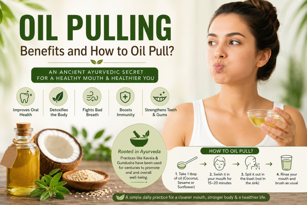
OIL PULLING – Benefits and How to Oil Pull? It is a common belief that oral health serves as a gateway to general health. This implies that oral health significantly impacts the general health and wellbeing of an individual.  Dental diseases have detrimental effects on the functionality and quality of life of individuals. In addition, a strong relationship has been established between various oral and systemic diseases. In fact, the prevention and treatment of dental caries and periodontal disease have been shown to reduce the risk of diabetes and heart disease significantly. The use of oil pulling can be frequently found in ancient medical text and is supported by recent studies for its efficacy and long-term use for maintaining and improving oral health.  The oral cavity serves as a focal point of entry for pathogens into the systemic circulation. While the host immune system of a healthy individual prevents the body from virulent microorganisms, a breach in the physical barriers in the oral cavity may provide access to into the systemic circulation. Similarly, a lack of oral hygiene allows an increase in virulent microbial colonization of the oral biofilm. Therefore, mechanical and chemical means of controlling the quantity and quality (virulence) of the oral biofilm is important in preventing systemic diseases and particularly periodontal diseases such as gingivitis and periodontitis (1). Oil is pulling or oil swishing is the ayurvedic way of maintaining oral health and improving overall immune system. Oil pulling also acts as an excellent detoxifying agent in healing the body inside. It is a widely accepted fact that most of the diseases start because of the unhealthy mouth. Most of the chronic illness are directly related to an unhealthy mouth like gum disease or tooth decay. As per Ayurveda – Oil pulling is incredibly effective in brightening teeth, healing gums, preventing bad breath, quenching inflammation, and healing oral infections. For example – Brushing is contra indicated in the cases of mouth ulcer, fever, indigestion, those who have tendency to vomit, asthma, cough, thirst. Oil pulling can be used to clean the oral cavity in all these cases (2). Gundusha and Kavala Graha are two primary oral cleansing techniques used in Ayurveda; as a specialized therapy to treat as well as to prevent oral diseases. Gundusha involves filling the mouth completely with fluid so that gargling is impossible. In Gundush, the oral cavity is filled completely with liquid medicine, held for about 3-5 minutes, and then released. In Kavala Graha, a comfortable amount of fluid is retained with the mouth closed for about 3 minutes and then gargled. It is a simple rejuvenating treatment, which, when done routinely, enhances the senses, maintains clarity, brings about a feeling of freshness, and invigorates the mind. These oral cleansing techniques can also benefit bad breath, dry face, dull senses, exhaustion, anorexia, loss of taste, impaired vision, sore throat, and all kapha related imbalances (9). This article – highlights the importance of incorporating oil pulling as a component of daily oral hygiene which can significantly improve oral and general health. What is Oil Pulling? Oil pulling, in CAM (Complementary and Alternative Medicine), is a technique that involves vigorous swishing of oil in the mouth, to achieve oral and systemic health benefits, like the modern-day use of mouthwashes and oral rinses. It is a powerful detoxifying Ayurvedic technique that has recently become very popular as a CAM remedy for many different health ailments. In the Ayurvedic text “Charaka Samhita,” it is mentioned as Kavala or Gundusha and is claimed to cure about 30 systemic diseases ranging from headache, migraine to diabetes and asthma (1). It has been used for centuries for the treatment and prevention of various oral and systemic diseases, using edible oils derived from either sunflower, sesame, or coconut. Oil pulling has been used extensively as a traditional Indian folk remedy for many years to prevent tooth decay, oral malodor (bad breath), bleeding gums, dryness of throat, cracked lips and for strengthening teeth, gums and the jaw (1).  In addition to brushing your teeth, flossing, and scraping your tongue, oil pulling with sesame or coconut oil is a safe and effective bonus to a healthy oral hygiene routine. It helps in rebalancing oral microbiome and improving oral and dental health. Ayurveda advises oil gargling to purify the entire system; as it holds that each section of the tongue is connected to different organ such as to the kidneys, lungs, liver, heart, small intestines, stomach, colon, and spine, similarly to reflexology and TCM (9).  Scientific evidence suggests that oil pulling therapy may reduce the total oral bacterial count and reduce plaque and gingival scores. Furthermore, it has also shown to diminish the susceptibility to dental caries from marked to slight or moderate level (1).  How does Oil Pulling work? (Mechanism of action) Both Western medicine and Ayurveda use the tongue as an important diagnostic tool, indicating that a healthy mouth and a healthy tongue are interrelated with the health of the entire body. Thus, supporting our oral hygiene and is a benefit for both our dental and general health (3). In fact, our mouths host over 600 different species of bacteria that populate the teeth, tongue, soft tissues of the cheeks and palates, and our tonsils. The oral cavity further adjoins the esophagus, nasal passages, sinuses, and the intricate ear cavities. You can see why bacteria in the mouth is a big deal! (3) Many of these bacteria are necessary for a healthy oral microbiome, but some, such as Streptococcus mutans, can cause problems if left unchecked—tooth decay, bad breath, gingivitis, and strep throat, to name a few. Poor oral hygiene can allow harmful bacteria to flourish, leading to various oral health issues (1, 3). Bacteria are single-celled organisms, enclosed by a lipid membrane. These bacteria in the mouth are attracted to the lipid structure of the oil, pulled from the oral tissue by adhering to the fat molecules of the oil, then flushed away through the act of swishing oil and spitting it out. This process helps in

Blog

EXPLORING THE LINK BETWEEN FATTY LIVER AND SIBO

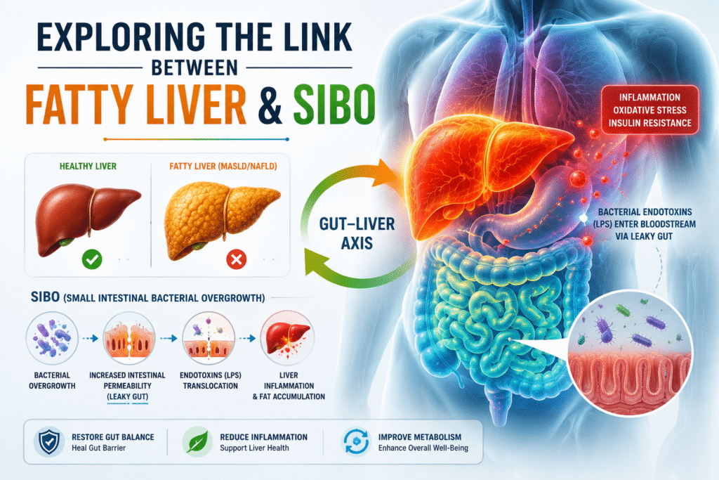
EXPLORING THE LINK BETWEEN FATTY LIVER AND SIBO Fatty liver disease, particularly Nonalcoholic Fatty Liver Disease (NAFLD), now known as Metabolic Dysfunction-Associated Steatotic Liver Disease (MASLD), has become a major global health concern. In recent years, MASLD has been the second leading cause of end-stage liver disease worldwide (28).  Increasing evidence reveals a fascinating connection between NAFLD and gut health, specifically a condition known as Small Intestinal Bacterial Overgrowth (SIBO). The correlation between small intestinal bacterial overgrowth (SIBO) and nonalcoholic fatty liver disease (NAFLD) has gained heightened acknowledgment, especially in the late phases of liver disease. Today, we explore the scientific links between these two conditions and why understanding this relationship matters. Understanding The Gut-Liver Axis The gut epithelium is a natural barrier that selects entry of useful substances present in the lumen, as nutrients, and keeps at bay bacteria, their bio-products and other potentially harmful elements. Tight junctions, specialized intercellular structures, assist this control. Derangement of the homeostasis between bacteria and the host, as occurs in SIBO (enhanced amount and/or changes in the type of bacteria in the gastrointestinal tract), may cause disruption of the intercellular tight junctions and subsequent increase in intestinal permeability leading to bacterial translocation (BT), i.e., transportation of bacteria and bacterial products from the intestinal lumen into the blood (4). The portal vein and the hepatic artery supply blood to the liver. The portal blood contains products of digestion and microbial products derived from the gut microbiota. This blood is carried to the liver. Therefore, Liver is the first site of exposure and filtration that consists of microbial products from the gut, such as LPS, lipopeptides, unmethylated DNA, and double-stranded RNA, which may evoke inflammatory reaction contributing to the progression of the liver disorder (4). This bidirectional relationship of the gut ecosystem and liver is imperative both physiologically and pathologically. Generally, the liver receives rich nutrients, microbial metabolites, and subproducts from the intestine and secretes bile into the small intestine (5). An integrated gut barrier also protects against toxins to maintain internal homeostasis.  This gut-liver axis is regulated and stabilized by a complex network of metabolic, immune, and neurosecretory interactions between the gut, microbiota, and liver. Disruption of this equilibrium may lead to gut dysbiosis and liver injury (5). SIBO as we know is the clinical manifestation of gut microbial dysbiosis. Therefore, the bidirectional relationship between Small Intestinal bacterial overgrowth (SIBO) and fatty liver disease, particularly non-alcoholic fatty liver disease (NAFLD), is characterized by mutual influences through gut-liver axis dysfunction, inflammation, and metabolic disturbances.  Association between SIBO and Fatty Liver Small intestinal bacterial overgrowth (SIBO) is a condition marked by excessive growth of microbes in the small intestine, resulting in various digestive issues including bloating, satiety, and malabsorption. In healthy people, the small bowel has a relatively low bacterial concentration, around 103–104 colony-forming units per milliliter (CFU/mL) (1,6). However, when this balance is disrupted, bacteria from the colon or oral cavity can colonize the small intestine, resulting in SIBO. Factors contributing to this condition include reduced gastric acid production, impaired intestinal motility, insufficient production of bile and dysfunction of the ileocecal valve (1,7). It may present in a range of symptoms, from moderate pain to severe nutritional deficiencies, weight loss, and shortages in crucial minerals and vitamins, including vit B12, A, D, E, iron, choline, calcium, fats, carbohydrates, protein and bile salt deconjugation (1,8). However, it has been shown that intestinal dysbiosis, endotoxemia (bacterial toxins in blood) and bacterial translocation may contribute to inflammation and Insulin Resistance (3,9,10,11,12). This directly seems to disrupt the functioning of the gut–liver axis, which may influence the incidence and progression of NAFLD (3,13). Non-alcoholic fatty liver Disease is the most frequent cause of chronic liver sickness globally, with a spectrum spanning from simple steatosis to inflammation of the hepatocytes, fibrosis, cirrhosis, and even hepatocellular carcinoma (1,14). The link between SIBO and nonalcoholic fatty liver disease (NAFLD) has attracted increased attention since studies show that the gut-liver axis plays a significant role in the pathophysiology of steatosis liver disease (1,15). The transfer of bacterial metabolites from the stomach to the liver may promote scarring and inflammation, thereby aggravating liver damage (1,16).  How SIBO affects Fatty Liver? 1.Inflammation – SIBO leads to an overgrowth of bacteria in the small intestine, causing increased intestinal permeability “known as leaky  gut or gut barrier dysfunction.” This allows bacterial endotoxins, especially lipopolysaccharides (LPS), to enter the bloodstream and reach  the liver via the portal vein, triggering chronic liver inflammation. These endotoxins and bacterial products activate inflammatory pathways (immune responses) in the liver, releasing proinflammatory cytokines (e.g. TNF-α, IL-6) which promote fat accumulation, insulin resistance, liver inflammation, fibrosis, and disease progression in NAFLD. Example – It has been demonstrated in animal models that a four-week HFD (high fat diet) increases LPS contained in the gut microbiota and plasma LPS concentrations two to three times, which is considered metabolic endotoxemia. The induction of metabolic endotoxemia in mice, by continuous subcutaneous infusion of LPS for four weeks, was followed by a rise in the following parameters: fasting glycemia, insulinemia, markers of inflammation, liver triglyceride content, liver insulin resistance, and whole body, liver and adipose tissue weight gain in a similar amount as occurred in HFD fed mice (4,17).  Large amount of fructose consumption is also related to increase in endotoxin serum levels, proinflammatory response and steatosis. It was demonstrated in an elegant study conducted by Bergheim et al. (4,18) that mice fed with fructose showed increased endotoxin levels in the portal blood, and higher intrahepatic lipid accumulation, lipid peroxidation and TNF-α expression.   2.Metabolic disruption – SIBO also disrupts bile acid metabolism, nutrient absorption, and adipose tissue function, contributing further to liver fat deposition and dysfunction.  Bile acids are synthesized in the liver from cholesterol, conjugated (joined) with glycine or taurine, and secreted into the small intestine, where they help digest fats and maintain gut microbial balance through their antimicrobial properties (3). In a healthy gut, most bile acids are absorbed in the ileum, while the rest reach the colon, where

Scroll to Top